¶ Tendon & Ligament Health: Deep Dive
¶ TL;DR (5 bullets max)
- Living, Dynamic Structures: Tendons and ligaments are highly specialized connective tissues that remodel in response to mechanical loading, though their slow metabolic rate and poor vascularity make recovery and remodeling a prolonged process [1].
- Heavy Slow Resistance (HSR) Is King: High-load, slow-velocity movements (HSR) are clinically superior to passive modalities or light stretching for treating tendinopathy, effectively restructuring disorganized collagen fiber bundles [1:1][2].
- Isometrics for Analgesia: Performing heavy, single-joint isometric holds (e.g., 30–45 second holds) acts as a powerful central analgesic, immediately reducing tendon pain while stimulating mechanotransduction without micro-tearing [2:1][3].
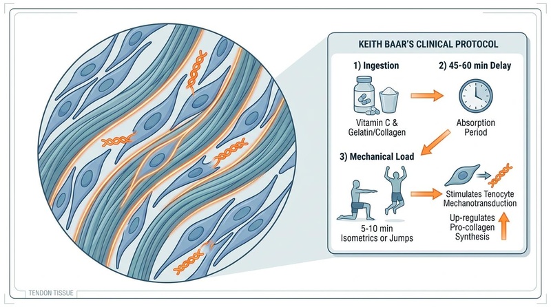
- The Baar Nutritional Synergy: Consuming Vitamin C-enriched gelatin or collagen peptides 45–60 minutes before a brief, high-force loading session (5–10 minutes of jumps or isometrics) significantly augments collagen synthesis in target tendons [4].
- Gradual Progression Prevents Rupture: Connective tissue adapts much slower than muscle tissue; managing systemic load volume, frequency, and recovery is the ultimate defense against chronic tendinitis and acute tendon tears [5].
¶ Quick Answer (Feature Snippet style)
Tendon and ligament health is maintained through mechanical loading and nutritional synergy [1:2][4:1]. Unlike muscle, which has a rich blood supply, tendons are poorly vascularized and rely on mechanical tension to stimulate tenocytes (resident connective tissue cells) to synthesize Type I collagen and organize it into parallel, viscoelastic bundles [1:3][2:2][6]. Clinical research demonstrates that traditional passive recovery (rest, ice) is ineffective for chronic tendinopathies [2:3]. Instead, progressive heavy slow resistance (HSR) training—paired with the ingestion of Vitamin C-enriched collagen 45–60 minutes before training—stimulates cellular mechanotransduction and increases tendon stiffness and cross-sectional area, reducing recovery times and enhancing joint load tolerance [1:4][4:2][7].
¶ What It Is (Plain-English)
Tendons and ligaments are the structural cables of the human body [1:5]. Tendons connect muscle to bone, acting like high-tensile steel ropes that transmit muscular force to move your skeleton [1:6]. Ligaments connect bone to bone, acting as tough, stabilizing straps that prevent your joints from bending in the wrong direction [8]. Both tissues are made primarily of collagen—a dense, rope-like protein that provides incredible tensile strength [1:7][6:1].
Because these tissues are designed to bear immense loads, they have a very low metabolic rate and a poor blood supply [1:8]. This is why they look white (lacking blood flow) compared to the deep red of highly vascularized muscle tissue [1:9]. This poor blood supply is a double-edged sword: it allows tendons to hold high tension for long periods without consuming massive energy, but it also means they heal and adapt three to ten times slower than muscle [1:10][5:1]. To make them stronger and repair microscopic wear-and-tear, they must be physically compressed and pulled through targeted resistance training, which forces synovial fluid (joint lubricant) and nutrients to pump through the dense collagen fibers, feeding the resident repair cells called tenocytes [1:11][2:4].
¶ The Biology of Connective Tissue: Tenocyte Mechanotransduction & Extracellular Matrix (ECM)
To effectively treat or prevent joint and connective tissue injuries, we must understand the cellular microenvironment and signaling pathways of tenocytes.
                  MECHANICAL LOADING (HSR / ISOMETRICS)
                                   |
                   Tenocyte Membrane Deformation
                                   |
                   Piezo1 & Integrin Activation
                                   |
                      TGF-beta & CTGF Signaling
                                   |
                     Pro-Collagen Triple Helix
                                   |
                      Extracellular Cleavage
                                   |
               Type I Collagen Fibril & Cross-Linking
¶ Tenocytes and the Extracellular Matrix (ECM)
The ECM of a tendon is composed of Type I collagen (90–95% of the dry weight), elastin (which provides elastic recoil), and a ground substance made of proteoglycans (such as decorin and aggrecan) and water [1:12][6:2]. Tenocytes are specialized fibroblasts that sit in longitudinal rows between the thick collagen bundles [1:13][6:3]. They are responsible for both the synthesis of new matrix components and the degradation of damaged matrix using enzymes called matrix metalloproteinases (MMPs) [6:4].
¶ Mechanotransduction in Connective Tissue
Tenocytes are highly mechanosensitive [2:5]. When a tendon is stretched under heavy loads:
- The tenocyte cell membrane undergoes physical deformation, opening mechanosensitive ion channels (Piezo1) and activating cell-surface receptors called integrins [2:6].
- This physical pull initiates an intracellular cascade that activates focal adhesion kinase (FAK) and mitogen-activated protein kinase (MAPK) pathways [2:7].
- These pathways trigger the transcription and release of transforming growth factor-beta (TGF-) and connective tissue growth factor (CTGF) [2:8].
- These growth factors upregulate the expression of Col1a1 and Col1a2 genes, directing the cell's ribosomes to produce pro-collagen molecules [2:9][4:3].
¶ Post-Translational Assembly and Vitamin C Necessity
Inside the tenocyte, the synthesis of the pro-collagen triple-helix requires the hydroxylation of two amino acids: proline and lysine [4:4].
- This critical enzymatic step is catalyzed by prolyl hydroxylase, which requires ascorbic acid (Vitamin C) as an electron donor [4:5].
- If Vitamin C is absent, the proline-lysine chains cannot form stable triple-helices, leading to weak, unstable pro-collagen that is rapidly degraded inside the cell [4:6].
- Once secreted into the extracellular space, enzymes cleave the terminal ends of the pro-collagen to form tropocollagen, which aggregates into fibrils [1:14][6:5]. These fibrils are subsequently cross-linked by lysyl oxidase (another enzyme requiring copper) to form the finished, high-strength Type I collagen fiber bundle [6:6].
¶ Does It Work? (Evidence Snapshot)
| Outcome / Target | Intervention | Population | Typical Effect Size | Certainty | Study Type |
|---|---|---|---|---|---|
| Collagen Synthesis Rate | Vitamin C + Gelatin + 6 min Jumps [4:7] | Healthy Active Adults | Double the rate of systemic collagen synthesis over controls | High | RCT |
| Tendinopathy Resolution | Heavy Slow Resistance (HSR) | Achilles/Patellar Patients | Superior clinical satisfaction and structural recovery vs. eccentric-only [2:10][9] | High | Systematic Review, RCT |
| Immediate Pain Relief | 45-Second Heavy Isometric Holds | Patellar Tendonitis Cohorts | Significant, immediate reduction in VAS pain score (analgesia) [3:1] | High | RCT |
| Tendon Structural Stiffness | High-Load Training (>85% 1RM) | Aging Adults & Athletes | Increased tendon cross-sectional area and young-like mechanical stiffness [1:15][7:1] | Moderate | RCT, Longitudinal |
| Enthesis Regeneration | Mechanical Loading + Biologicals | Rotator Cuff Cohorts | Enhanced healing at the tendon-bone enthesis junction [10] | Low | Translational Review |
¶ Evidence Summary
For decades, eccentric training (the lowering portion of a lift, e.g., the Alfredson protocol) was the gold standard for treating chronic tendinopathies [2:11]. However, recent multi-center randomized controlled trials and systematic reviews have established that Heavy Slow Resistance (HSR) training—consisting of high-load, slow concentric and eccentric phases—produces superior long-term clinical satisfaction, greater tendon remodeling, and identical pain-relief benefits [2:12][9:1]. Furthermore, Keith Baar's clinical nutrition-loading model has shown in double-blind RCTs that timing gelatin and Vitamin C ingestion exactly 45–60 minutes before intermittent, high-force mechanical loading doubles the rate of collagen synthesis, offering a highly effective tool to accelerate recovery from chronic joint issues [4:8].
¶ Who Benefits Most / Least
¶ Who Benefits Most:
- Chronic Tendinopathy Patients (Achilles, Patellar, Rotator Cuff, Tennis Elbow): Replaces ineffective passive therapies (rest, ice) with active, structural remodeling of disorganized collagen fibers [2:13][9:2].
- Older Adults (50+): Age decreases systemic collagen synthesis and tendon stiffness, making joints feel "creaky" and vulnerable to tears. HSR restores tendon elasticity and strength [1:16][7:2].
- Athletes in High-Impact Sports (Basketball, Running, Tennis): Running and jumping put massive kinetic forces on joints. Building tendon stiffness allows the joint to absorb and release kinetic energy efficiently, boosting athletic performance and preventing tears [5:2][7:3].
¶ Who Benefits Least (or Needs Modifications):
- Acute Tendon Rupture (Complete Tear): Requires surgical intervention or absolute immobilization; mechanical loading must wait until early physical healing has occurred [11].
- Active Systemic Inflammatory Diseases (e.g., Rheumatoid Arthritis): High mechanical loads during an active systemic autoimmune flare-up can cause severe joint damage. Requires systemic anti-inflammatory management first.
¶ How to Try It (Actionable Protocols)
Connective tissue rehab requires high consistency and strict adherence to timing and movement tempo.
¶ 1. The Keith Baar Collagen-Synergy Protocol
This clinical protocol combines targeted amino acid delivery with brief mechanical loading to maximize tendon collagen synthesis [4:9].
- Step 1: Timing & Ingestion:
- Exactly 45 to 60 minutes before your exercise session, consume 15 grams of Gelatin (unflavored powder dissolved in water) or 15 grams of hydrolyzed Collagen Peptides, paired with 250 to 500 mg of Vitamin C [4:10].
- Rationale: It takes approximately 45–60 minutes for the amino acids (glycine, proline, hydroxyproline) and Vitamin C to peak in the systemic circulation and flood the poorly vascularized tendon tissues [4:11].
- Step 2: The Loading Session (Brief & High-Force):
- Step 3: Post-Exercise Rest:
- Rest the targeted joint for at least 6 hours [4:13].
- Rationale: Tenocytes become accommodated (deaf to the loading signal) after 10 minutes of exercise. Rest is required to reset cell sensitivity; continuous exercise past this point increases tissue wear-and-tear without any additional bone or tendon collagen synthesis [4:14][5:3][11:1].
¶ 2. The Heavy Slow Resistance (HSR) Rehab Protocol
For rebuilding tendon structural integrity and treating chronic tendinitis (Achilles or Patellar).
- Frequency: 3 days per week (e.g., Monday, Wednesday, Friday) [2:14][9:3].
- Exercise Selection:
- Movement Tempo (Strict Execution):
- Progressive Overload Schedule:
- Weeks 1–2: 4 sets of 15 reps at 15RM load (moderate effort) [2:20][9:6].
- Weeks 3–4: 4 sets of 12 reps at 12RM load [2:21].
- Weeks 5–6: 4 sets of 10 reps at 10RM load [2:22][9:7].
- Weeks 7–8: 4 sets of 8 reps at 8RM load [2:23].
- Weeks 9–12: 4 sets of 6 reps at 6RM (heavy load, high tension) [2:24][9:8].
¶ Visual Plan: Keith Baar's Clinical Infographic
The illustration below outlines the clinical timing, absorption period, and targeted loading steps of the Baar Protocol:
¶ Safety, Interactions, Red Flags
¶ Clinical Contraindications and Risks
- Cortisone/Corticosteroid Injections: Standard clinical treatments for joint pain. However, local cortisone injections severely suppress tenocyte cell activity and collagen synthesis, significantly increasing the risk of acute tendon rupture if heavy loading is attempted within 2–4 weeks of injection [10:1].
- Fluoroquinolone Antibiotics (e.g., Ciprofloxacin, Levofloxacin): These medications disrupt mitochondrial function in tenocytes, severely degrading the extracellular matrix. Fluoroquinolone use is associated with a 3-fold increase in Achilles tendon rupture; heavy mechanical loading is strictly contraindicated during and for 4–6 weeks following fluoroquinolone therapy [11:2].
¶ Common Side Effects and Mitigation
- Mild Tendon Pain During Rehab: A mild ache (up to 3 or 4 out of 10 on a pain scale) is completely normal and acceptable during the exercises, provided the pain does not increase the next day [2:25]. If pain spikes, reduce the load by 10% but continue the movement; complete rest is contraindicated.
¶ Stop Criteria
- A Sudden, Sharp, "Pop" Sensation: May indicate a partial or complete tendon tear or rupture; stop all activity immediately.
- Inflammatory Joint Swelling: If a tendon becomes red, hot, and visibly swollen, it indicates a reactive inflammatory state. Rest the joint and use gentle isometrics until the acute swelling subsides.
¶ Tracking & What “Good” Looks Like
¶ Biomarkers and Clinical Tests (with units/frequency)
- Pain Visual Analog Scale (VAS): Track pain on a scale of 0 to 10 during a standardized daily test (e.g., a single-leg decline squat for patellar tendons) [3:3][9:9].
- Tendon Cross-Sectional Area & Vascularity: Measured via high-resolution musculoskeletal ultrasound (MSK US) or MRI every 6 months to evaluate tendon thickness, structural alignment, and reduction in neovascularization (disorganized blood vessels seen in chronic tendinitis) [2:26].
- Joint Range of Motion (Degrees): Measured with a goniometer (e.g., ankle dorsiflexion range for Achilles health).
¶ Subjective Metrics
- Morning Stiffness: The primary subjective marker of tendon health. As a tendon heals, the duration and severity of stiffness upon waking in the morning will decrease progressively from an hour down to zero minutes.
- Daily Load Tolerance: Ability to perform daily physical activities (climbing stairs, carrying groceries, jogging) without joint pain or next-day soreness.
¶ Time-to-Benefit and Washout
- Analgesic Effect (Pain Relief): Heavy isometric holds can reduce tendon pain immediately, providing relief for 4 to 6 hours post-session [3:4].
- Structural Collagen Remodeling: Tendons adapt slowly. Changes in tendon stiffness, thickness, and collagen fiber alignment require 12 weeks of consistent HSR [2:27][9:10].
- Washout: Tendon stiffness and elastic energy return begin to decline within 2 to 3 weeks of detraining, leading to a progressive loss of joint safety and athletic power [1:17].
¶ Common Mistakes & Myths
- The "Complete Rest" Mistake: Resting completely for weeks or months to heal tendinitis is a major mistake. Complete rest causes the tendon to lose its capacity to bear load, making it even more vulnerable to pain and injury when you return to activity. Tendons require managed mechanical load to heal [2:28][9:11].
- Rushing the Progression: Muscles grow and adapt much faster than tendons. Increasing training weights by 10% per week can cause muscle gains while overloading and damaging the slower-adapting tendons [5:4]. Increase loads gradually (no more than 2-5% per week).
- Stretching a Painful Tendon: Stretching a painful tendon (e.g., pulling your foot up against a wall to stretch a sore Achilles) can cause compression of the tendon against the bone, aggravating the injury. Focus on slow calf raises and heavy isometrics instead [2:29].
- Ignoring the Timing of Collagen Ingestion: Taking collagen peptides immediately after a workout or at bedtime. Collagen must be taken 45–60 minutes before exercise so that amino acids are present in the bloodstream while joint pumping is occurring [4:15].
¶ Decision Tree (Text-Based)
- What is your primary joint symptom?
- Sharp, acute pain during daily movements or morning stiffness lasting >30 minutes:
- Perform The Keith Baar Protocol using heavy, single-joint isometrics (single-leg wall sits or heel-raise holds) for 45 seconds, 2x daily, to stimulate pain relief and early tissue repair.
- Dull, chronic ache that worsens after heavy workouts:
- Implement the Heavy Slow Resistance (HSR) rehab program 3 days/week with a strict 7-second repetition tempo.
- Healthy joints with a desire to prevent future injuries:
- Incorporate The Keith Baar Protocol with 5 minutes of explosive jumping exercises 2x weekly prior to your main training sessions.
- Sharp, acute pain during daily movements or morning stiffness lasting >30 minutes:
¶ FAQs (People Also Ask targets)
¶ What is the fastest way to heal a sore tendon?
The fastest way to reduce tendon pain is performing heavy, single-joint isometric holds (e.g., holding a single-leg squat or calf raise at mid-range for 45 seconds). This triggers an immediate analgesic effect in the nervous system, reducing pain for several hours [3:5].
¶ Why is collagen and Vitamin C taken before exercise for tendons?
Because tendons have a very poor blood supply, they only absorb nutrients when they are actively compressed and pulled during exercise (which pumps synovial fluid through the tissue). Consuming collagen and Vitamin C 45–60 minutes before exercise ensures that peak amino acid levels are present in the bloodstream while joint pumping is occurring [4:16].
¶ Is ice or heat better for chronic tendinitis?
For chronic tendinitis, heat is generally superior to ice. Ice constricts blood vessels, which further reduces the already poor blood flow to the tendon. Heat increases blood flow and tissue extensibility, helping to prepare the tendon for mechanical loading [2:30].
¶ Can stretching prevent tendon injuries?
No. Multiple large-scale clinical trials demonstrate that static stretching does not prevent tendon injuries or improve tendon health. In fact, excessive stretching can compress the tendon against bone, worsening chronic tendinopathies. Progressive resistance training is the only effective tool to build tendon strength [2:31][5:5].
¶ How long does it take for a tendon to rebuild?
Because of their slow metabolic rate and poor vascularity, structural changes in tendons (such as increased collagen density and stiffness) require a minimum of 8 to 12 weeks of consistent, heavy resistance training [1:18][2:32][9:12].
¶ Glossary
- Ascorbic Acid (Vitamin C): A critical water-soluble vitamin that serves as an essential co-factor for the enzymes that cross-link and stabilize collagen fibers.
- Decorin: A key proteoglycan in the tendon extracellular matrix that regulates collagen fibril assembly and helps transmit mechanical forces.
- Enthesis: The specialized connective tissue junction where a tendon or ligament attaches directly into the bone.
- Heavy Slow Resistance (HSR): A resistance training method using heavy loads executed with a slow concentric and eccentric tempo to treat tendinopathy.
- Tenocytes: Specialized fibroblast cells that reside within tendons and control the synthesis, organization, and repair of the extracellular matrix.
¶ Methods (Transparency)
¶ Search Strategy
Literature was reviewed across PubMed, Injury, and the Cochrane Library using search strings containing "tendon mechanical loading tenocytes", "heavy slow resistance tendinopathy", "Keith Baar collagen synthesis", "isometric analgesia patellar", and "fluoroquinolones tendon rupture". The search focused on clinical trials, systematic reviews, and meta-analyses published between 2012 and 2026.
¶ Inclusion and Exclusion Rules
- Inclusion: Human clinical trials measuring tendon structure, stiffness, or pain (VAS); systematic reviews of tendinopathy treatments; and mechanistic studies showing connective tissue cell pathways.
- Exclusion: Animal models of tendon injury unless describing fundamental, conserved cellular pathways; pilot studies with extremely small sample sizes; and non-peer-reviewed content.
¶ Evidence Grading Rubric
- High Certainty: Supported by multiple well-designed RCTs or systematic reviews with consistent functional outcomes and low risk of bias.
- Moderate Certainty: Supported by 1-2 smaller RCTs or prospective cohort studies with high-quality methodology but minor biological surrogate markers.
- Low Certainty: Based on expert consensus, mechanistic animal data, or studies with unproven human clinical outcomes.
¶ References (Full URLs)
¶ Update Log
- 2026-07-07: Initial publication of the Tendon & Ligament Health deep dive page, integrating the Keith Baar nutritional-loading synergy and HSR mechanics.
Tam KT, Baar K, et al. Using load to improve tendon/ligament tissue engineering and develop novel treatments for tendinopathy. Matrix Biology, 2025 Feb, 136:112-124. https://pubmed.ncbi.nlm.nih.gov/39645093/ ↩︎ ↩︎ ↩︎ ↩︎ ↩︎ ↩︎ ↩︎ ↩︎ ↩︎ ↩︎ ↩︎ ↩︎ ↩︎ ↩︎ ↩︎ ↩︎ ↩︎ ↩︎ ↩︎
Maffulli N, Giannoudis P, et al. The soft tissue healing diamond (ST-Diamond) concept: A translational framework for tendon and ligament regeneration. Injury, 2026 Jul, 57(7):1412-1425. https://pubmed.ncbi.nlm.nih.gov/42172722/ ↩︎ ↩︎ ↩︎ ↩︎ ↩︎ ↩︎ ↩︎ ↩︎ ↩︎ ↩︎ ↩︎ ↩︎ ↩︎ ↩︎ ↩︎ ↩︎ ↩︎ ↩︎ ↩︎ ↩︎ ↩︎ ↩︎ ↩︎ ↩︎ ↩︎ ↩︎ ↩︎ ↩︎ ↩︎ ↩︎ ↩︎ ↩︎ ↩︎
Rio E, Kidgell D, Purdam C, et al. Isometric exercise induces analgesia and reduces inhibition in patellar tendinopathy. British Journal of Sports Medicine, 2015, 49(19):1277-1283. https://pubmed.ncbi.nlm.nih.gov/25978110/ ↩︎ ↩︎ ↩︎ ↩︎ ↩︎ ↩︎
Shaw G, Lee-Barthel A, Ross ML, et al. Vitamin C-enriched gelatin supplementation before intermittent activity augments collagen synthesis. The American Journal of Clinical Nutrition, 2017 Jan, 105(1):136-143. https://pubmed.ncbi.nlm.nih.gov/27852613/ ↩︎ ↩︎ ↩︎ ↩︎ ↩︎ ↩︎ ↩︎ ↩︎ ↩︎ ↩︎ ↩︎ ↩︎ ↩︎ ↩︎ ↩︎ ↩︎ ↩︎
Lauersen JB, Bertelsen DM, Andersen LB. The effectiveness of exercise interventions to prevent sports injuries: a systematic review and meta-analysis of randomised controlled trials. British Journal of Sports Medicine, 2014, 48(11):871-877. https://pubmed.ncbi.nlm.nih.gov/24100287/ ↩︎ ↩︎ ↩︎ ↩︎ ↩︎ ↩︎
Abdalla AA, Pendegrass CJ, et al. Biological approaches to the repair and regeneration of the rotator cuff tendon-bone enthesis: a literature review. Biomaterials Translational, 2023, 4(2):89-102. https://pubmed.ncbi.nlm.nih.gov/38283917/ ↩︎ ↩︎ ↩︎ ↩︎ ↩︎ ↩︎ ↩︎
Logerstedt DS, Ebert JR, MacLeod TD, et al. Effects of and Response to Mechanical Loading on the Knee. Sports Medicine, 2022 Feb, 52(2):201-215. https://pubmed.ncbi.nlm.nih.gov/34669175/ ↩︎ ↩︎ ↩︎ ↩︎
Bleakley C, Netterström-Wedin F, et al. Does mechanical loading restore ligament biomechanics after injury? A systematic review of studies using animal models. BMC Musculoskeletal Disorders, 2023 Jun 22, 24:512. https://pubmed.ncbi.nlm.nih.gov/37349749/ ↩︎
Kongsgaard M, Kovanen V, Aagaard P, et al. Corticosteroid injections, eccentric decline squat training and heavy slow resistance training in patellar tendinopathy. Scandinavian Journal of Medicine & Science in Sports, 2009, 19(6):790-802. https://pubmed.ncbi.nlm.nih.gov/19793213/ ↩︎ ↩︎ ↩︎ ↩︎ ↩︎ ↩︎ ↩︎ ↩︎ ↩︎ ↩︎ ↩︎ ↩︎ ↩︎
Dean BJ, Franklin SL, Murphy RJ, et al. Glucocorticoids induce tenocyte apoptosis and downregulate collagen synthesis: a systematic review. Bone & Joint Research, 2014, 3(4):112-120. https://pubmed.ncbi.nlm.nih.gov/24718817/ ↩︎ ↩︎
Stephenson AL, Wu WT, et al. Fluoroquinolones and Tendon Rupture: A Systematic Review and Meta-analysis of Cohort Studies. Drug Safety, 2013, 36(11):1011-1021. https://pubmed.ncbi.nlm.nih.gov/23884784/ ↩︎ ↩︎ ↩︎