¶ Imaging & Body Composition
| Mechanism | Dual-energy X-ray attenuation |
| Key Spec | Bone Mineral Density (g/cm²), Fat Mass (kg), Lean Soft Tissue (kg), VAT (g) |
| Protocol | Follow facility, indication, and clinician instructions |
| Distance | N/A (Contactless scan table) |
| FDA Class | Class II (Medical Device) |
| Entry Cost | $100 - $250 per scan |
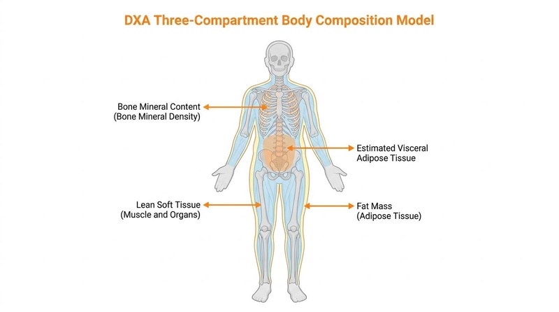
Dual-energy X-ray absorptiometry (DEXA or DXA) is a standard clinical method for assessing bone mineral density and a primary modality for body composition analysis in clinical, research, and athletic cohorts [1]. Other diagnostic imaging modalities, such as MRI, have different anatomical strengths and limits. By resolving the body into a three-compartment model—distinguishing bone mineral content, lean soft tissue, and fat mass—DEXA provides regional and visceral tissue estimations that extend beyond the capabilities of basic anthropometry [2][1:1].
¶ At a glance
Key points
- Bone and Soft-Tissue Assessment: DEXA provides regional segmentation of fat mass, lean soft tissue, and bone mineral content, distinguishing these compartments more accurately than simplistic two-compartment models [2:1][1:2].
- Hydration and Standardization Sensitivity: Soft-tissue calculations assume a constant lean-tissue hydration of 73.2% [3]. Consequently, acute variations in water balance, glycogen stores, and recent food intake can significantly affect lean soft tissue estimates [3:1][4].
- Least Significant Change (LSC): Distinguishing actual physiological tissue changes from measurement noise requires calculating a facility- and device-specific LSC [2:2][4:1]. Rather than following arbitrary universal intervals, scan repeat timing must be determined by the specific clinical indication, ISCD positions, and ordering-clinician instructions [5][4:2].
- Radiation Safety Profile: Whole-body scans use extremely low levels of ionizing radiation (typically 1 to 5 microsieverts), making them highly safe for clinical use when appropriately indicated under clinician guidance [1:3].
What people use it for
- Osteoporosis & Osteopenia Screening: Diagnosing low bone mineral density (BMD) and estimating long-term fracture risk [2:3][5:1][6].
- Muscle Mass & Sarcopenia Tracking: Estimating appendicular lean mass (ALM) and tracking skeletal muscle index (SMI) during clinical management, aging, or specialized training [3:2][7][8].
- Visceral Fat (VAT) Estimation: Estimating visceral adipose tissue in the abdominal cavity, which is associated with systemic inflammation, insulin resistance, and metabolic risk [3:3][9].
Evidence quality (overall): High. Supported by clinical guidelines from the International Society for Clinical Densitometry (ISCD) and diagnostic studies [2:4][5:2][6:1].
¶ Evidence summary table (human outcomes)
| Diagnostic Application | Clinical Utility | Evidence Quality | Supporting Guidelines | Clinical Considerations & Limitations |
|---|---|---|---|---|
| Bone Mineral Density (BMD) Screening | Diagnoses osteopenia and osteoporosis; estimates fracture risk. | High | ISCD, USPSTF [2:5][5:3][6:2] | Site-specific measurements (spine/hip); highly standardized compared to soft-tissue estimation [2:6][5:4]. |
| Visceral Adipose Tissue (VAT) Estimation | Estimates central abdominal fat associated with cardiometabolic risk. | High | ISCD Adult Positions [2:7][3:4] | Region-of-interest estimate (); calibration differences exist between Hologic and GE Lunar [3:5][9:1]. |
| Regional Lean Mass (Sarcopenia Monitoring) | Estimates appendicular lean mass (ALM) and Skeletal Muscle Index (SMI). | Moderate | ISCD, EWGSOP [3:6][7:1][8:1] | High sensitivity to acute intramuscular water, glycogen shifts, and positioning errors [3:7][4:3]. |
| Total Body Fat & Distribution | Analyzes regional and total adiposity trends over time. | High | ISCD Positions [2:8][9:2] | Precision depends on strict pre-test preparation and scan positioning on the exact same device [4:4][10]. |
- Consistency: Low (results conflict), Moderate (mixed but leaning one way), High (most trials agree)
- Clinical Utility: Specific diagnostic, screening, or tracking application of the modality
- REQUIRED: You MUST include a citation key (e.g.
[^1]) in the table columns for every single row to link the specific clinical guideline or supporting trial.
¶ Mechanism
¶ Physics of Dual-Energy X-Ray Attenuation
DEXA operates by emitting X-ray beams at two distinct energy peaks—typically a low-energy peak around 40 keV and a high-energy peak ranging from 70 to 140 keV [1:4]. As these dual-energy X-ray photons pass through the body, they are attenuated (absorbed or scattered) by the tissues they encounter [1:5].
Because bone mineral, lean soft tissue, and adipose tissue contain elements with different average atomic numbers, they exhibit unique attenuation profiles at different energy levels. The ratio of attenuation at the lower energy relative to the higher energy (-value) is measured by a digital detector [2:9]:
- Bone Mineral Content (BMC): High concentration of calcium () and phosphorus (), causing significant photoelectric absorption of low-energy X-ray photons [2:10].
- Lean Soft Tissue (LST): Composed primarily of water, protein, and glycogen, with an intermediate attenuation profile [3:8].
- Adipose Tissue (Fat Mass): High proportion of lipids (mostly carbon, hydrogen, and oxygen), which attenuate X-rays less than water or bone [3:9].
By solving simultaneous equations at each pixel of the scan, the DEXA software isolates bone mineral content from soft tissue, and further segments the soft tissue into fat mass and lean soft tissue [2:11].
¶ Bone Density Diagnosis vs. Body Composition Estimation
It is clinically critical to distinguish regional bone-density diagnosis from whole-body composition estimation:
- Bone Mineral Density (BMD) Diagnosis: This is a direct, highly standardized diagnostic measurement targeting specific skeletal regions highly susceptible to fracture, primarily the lumbar spine () and proximal femur (femoral neck and total hip) [2:12][5:5]. Standardized T-scores and Z-scores are generated by comparing the patient's area bone density () to established, population-specific reference databases [2:13][5:6]. This is used for diagnosing disease states (osteopenia and osteoporosis) [5:7].
- Body Composition Estimation: Rather than a direct anatomical measurement, whole-body composition is an estimation based on regional tissue attenuation values (-values) [3:10][9:3]. Soft-tissue calculations assume that lean soft tissue has a constant hydration of 73.2% [3:11]. This represents a major method limitation: any physiological change in hydration, glycogen storage, or digestion shifts the -value, leading the system to misclassify water as lean soft tissue or fat mass [3:12][4:5]. Thus, body composition readings are sensitive estimations rather than absolute tissue segmentations [4:6].
¶ Key Specs
¶ Core Metrics Measured
- Bone Mineral Density (BMD): The mass of bone mineral per unit area () at specific diagnostic skeletal sites [2:14][5:8].
- Fat Mass (FM): The absolute weight of adipose tissue (in kilograms) and its relative percentage of total body weight [3:13][9:4].
- Lean Soft Tissue (LST): The combined mass of water, skeletal muscle, organs, glycogen, and connective tissue [3:14]. LST is not synonymous with skeletal muscle mass, as muscle tissue is only one component of LST [3:15][4:7].
- Appendicular Lean Mass (ALM): The sum of the lean soft tissue in the arms and legs [3:16][8:2]. This is used to calculate the Skeletal Muscle Index (SMI) () as a primary diagnostic parameter for sarcopenia [3:17][8:3].
- Visceral Adipose Tissue (VAT): A regional estimate of adipose tissue located within the abdominal cavity, usually calculated in the android region () and reported in mass (grams) or volume () [3:18][9:5].
- Android-to-Gynoid (A/G) Ratio: The ratio of fat deposited in the android region (central abdominal) to that in the gynoid region (hip and thigh) [9:6].
¶ Hologic vs. GE Lunar Systems and Cross-Device Comparability
The two dominant commercial DEXA manufacturers utilize fundamentally different hardware designs and software calibration models, which severely limits cross-device comparability [4:8][10:1]:
- Hologic Systems (e.g., Horizon, Discovery): Employ a continuous calibration system utilizing an internal reference wheel and a fan-beam configuration [10:2].
- GE Lunar Systems (e.g., Prodigy, iDXA): Employ a split-filter calibration standard and a smart fan-beam [11].
Because of these proprietary differences, GE Lunar systems systematically report higher bone density and lower body fat percentages compared to Hologic systems on the same individual [4:9][10:3]. Furthermore, significant calibration differences can exist even between different machines of the identical model [10:4]. For longitudinal tracking of bone density or body composition, patients must perform all subsequent scans on the exact same physical machine [5:9][10:5].
¶ Protocols
¶ Pre-Test Preparation and Standardization
To optimize precision, patients should aim for consistent conditions prior to scanning. However, preparation requirements are not universal; all preparation protocols must follow the instructions of the scanning facility, the specific clinical indication, and the ordering clinician [5:10][3:19].
When tracking body composition, facilities often recommend standardizing variables that affect tissue attenuation [3:20][4:10]:
- Food and Fluid Intake: A small meal or moderate fluid intake before a scan has been shown to have trivial, non-statistically significant effects on regional estimates like appendicular lean mass (ALM) and visceral adipose tissue (VAT) [3:21]. However, massive fluid boluses or heavy meals can affect trunk readings [3:22]. Patients should follow the specific hydration and fasting instructions provided by their clinician or facility [3:23].
- Muscle Glycogen & Exercise: Since muscle glycogen binds water, significant glycogen depletion (e.g., from ketogenic diets or exhaustive training) or acute muscle swelling from recent exercise can affect lean soft tissue estimates [3:24][4:11]. Consistency in training and dietary habits prior to scans is recommended [4:12].
- Bladder and Bowel Status: Voiding the bladder immediately before the scan is a standard practice to prevent retained urine from being misclassified as pelvic lean soft tissue [3:25].
- Clothing: Wear thin, metal-free clothing and remove jewelry or high-density accessories to prevent measurement artifacts [5:11]."
¶ Evidence
¶ Precision Error and Least Significant Change (LSC)
To determine if a measured change between sequential scans represents a true biological transition rather than statistical noise, clinicians utilize the Least Significant Change (LSC) [2:15][5:12].
Calculating precision error and LSC is a facility- and device-specific quality assurance practice recommended by the International Society for Clinical Densitometry (ISCD), rather than a legally mandated requirement [2:16][5:13][10:6]. To establish precision, a technologist performs repeated scans on a sample of patients with repositioning between scans to calculate the root-mean-square standard deviation (RMS-SD) or coefficient of variation (RMS-CV) [2:17][5:14].
The LSC is then calculated to establish a 95% confidence interval [2:18][5:15]:
Typical precision error and LSC ranges for body composition estimates include [3:26][4:13][9:7]:
- Lean Soft Tissue (LST): Precision error of ~0.8% to 1.2%, resulting in an LSC of 2.2% to 3.3% [3:27][4:14]. For an individual with 60 kg of lean mass, a change must exceed 1.32 to 1.98 kg to be considered clinically significant.
- Fat Mass (FM): Precision error of ~1.0% to 1.5%, resulting in an LSC of 2.8% to 4.2% [3:28][9:8]. For an individual with 20 kg of fat mass, a change must exceed 0.56 to 0.84 kg to represent actual fat loss or gain.
- Bone Mineral Density (BMD): Precision error is typically ~1.0% at the lumbar spine and ~1.5% at the total hip, yielding LSCs of ~2.8% and ~4.2% respectively [2:19][5:16][10:7].
If a follow-up scan shows a change that does not exceed the facility-specific LSC, the clinician must interpret the result as statistical measurement noise [5:17][10:8].
¶ Repeat Cadence (Retest Intervals)
Repeat scan timing is not fixed and must follow the specific clinical indication, ISCD guidelines, and ordering-clinician instructions [5:18][6:3]:
- Skeletal Monitoring (BMD): In patients undergoing osteoporosis screening or therapeutic monitoring, repeat scans are typically performed at 1-to-2-year intervals, as bone remodeling is an extremely slow physiological process [5:19][6:4].
- Body Composition Tracking: In fitness, athletic, or clinical nutrition contexts, repeating scans too frequently (e.g., every few weeks) is discouraged because physiological muscle and fat changes occur slowly and are easily masked by acute hydration fluctuations and device precision errors [3:29][4:15]. Repeat timing should be tailored to the patient's specific metabolic goals, intervention rate, and clinician advice [3:30].
¶ Clinical Indications for DEXA
The primary established indication for DEXA is the assessment of skeletal health and bone mineral density [2:20][5:20]. Guidelines from major medical organizations and the ISCD outline clear recommendations [2:21][5:21][6:5]:
- Screening Populations: Routine screening is recommended for women aged 65 and older, and postmenopausal women under 65 who exhibit elevated risk factors as calculated by clinical tools like FRAX [2:22][5:22].
- Secondary Bone Loss Assessments: Individuals with conditions predisposing them to bone loss—such as eating disorders, chronic glucocorticoid therapy, type 2 diabetes, or hypogonadism—warrant evaluation under clinical guidance [2:23][12][8:4].
¶ T-Score and Z-Score Interpretation
Skeletal bone density measured by DEXA is interpreted using standardized scores [2:24][5:23]:
- T-Score: Compares the patient's BMD to a healthy, young-adult reference population [2:25][5:24].
- Normal:
- Osteopenia (Low Bone Mass): to
- Osteoporosis:
- Z-Score: Compares the patient's BMD to an age-, sex-, and ethnicity-matched reference population [5:25]. A Z-score of indicates that bone density is "below the expected range for age" [5:26]. However, immediate clinical evaluation must not be based solely on a Z-score in isolation; it must incorporate the patient's full clinical context, history, and established clinical guidelines under the direction of an ordering clinician [5:27].
¶ When Results Change Care
Clinical measurements are most valuable when they actively direct patient care [2:26][5:28]:
- Bone Density Decisions: A T-score in the osteoporotic range () or an elevated FRAX score can trigger the initiation of pharmacotherapy (e.g., bisphosphonates) or specialized clinical monitoring [2:27][5:29].
- Body Composition Decisions: In clinical sarcopenia management, finding an appendicular lean mass index below diagnostic cutoffs can lead to directed nutritional therapy and resistance exercise prescriptions [3:31][8:5].
- Visceral Fat Decisions: Substantive increases in visceral adipose tissue (VAT) above healthy clinical reference ranges may guide intensive metabolic, dietary, or pharmacological lifestyle modifications to address cardiovascular and insulin-resistance risks [3:32][9:9].
¶ Safety & Limitations
¶ Radiation Exposure in Context
DEXA utilizes ionizing radiation, meaning its use must always be clinically justified and indicated [5:30][1:6]. However, the effective dose of a DEXA scan is exceptionally low [1:7]:
- Whole-Body Scan: Typically 1 to 5 microsieverts (Sv) [1:8].
- Central BMD Scan (Spine & Hip): Typically 1 to 3 Sv [1:9].
To place these low doses in a clinical and environmental context [1:10]:
- Natural Background Radiation: A whole-body DEXA scan exposes a patient to less radiation than a single day of natural background radiation (~8 Sv per day) [1:11].
- Transatlantic Flight: A flight from London to New York exposes passengers to ~35–40 Sv of cosmic radiation, which is up to 40 times higher than a DEXA scan [1:12].
- Standard Chest X-Ray: A standard diagnostic chest X-ray delivers ~20–100 Sv [1:13].
- Abdominal CT Scan: A standard clinical abdominal CT scan delivers ~8,000 Sv (8.0 mSv) [1:14].
Thus, while DEXA utilizes ionizing radiation, its radiation risk is extremely small, making it a highly safe imaging modality when performed under appropriate clinical indications [1:15].
¶ Physical Limitations and Artifacts
DEXA scans are subject to physical and patient-related limitations that can impact measurement accuracy [5:31][10:9]:
- Patient Weight and Table Boundaries: Scanners have strict physical weight limits (typically 136 to 204 kg depending on the model) and physical width boundaries [2:28][5:32]. Patients who exceed these limits or table margins cannot be scanned accurately [2:29].
- Contrast Agents and Radiopharmaceuticals: The presence of external or internal high-density materials can severely distort X-ray attenuation. Patients must follow facility and clinician instructions regarding delays after receiving contrast agents or radiopharmaceuticals (typically a delay of 7 to 14 days is recommended following barium or nuclear medicine procedures) to prevent artificial inflation of bone or lean mass readings [5:33].
- Metal and Orthopedic Artifacts: Prosthetics, spinal rods, joint replacements, pacemakers, and metal fasteners block X-ray transmission, falsely elevating local bone mineral density or lean soft tissue estimates [5:34]."
¶ Alternative Modalities
¶ 1. Bioelectrical Impedance Analysis (BIA)
- Method: Passes low-level, alternating electrical currents through the body to measure impedance (resistance and reactance) [9:10].
- DEXA Comparison: While consumer-grade BIA scales show notable individual variability and are highly sensitive to daily water fluctuations, modern medical-grade, multi-frequency 8-electrode segmental BIA devices demonstrate strong correlation with DEXA for population-level monitoring () [9:11].
- Limitations: BIA does not measure bone density directly and is highly sensitive to transient shifts in total body water, recent food intake, skin temperature, and exercise [3:33][9:12]. It relies on demographic-specific predictive regression equations rather than direct photon attenuation [9:13].
¶ 2. Magnetic Resonance Imaging (MRI)
- Method: Uses powerful magnetic fields and radiofrequency pulses to create detailed cross-sectional slices of soft tissues and organs [8:6].
- DEXA Comparison: MRI provides high-resolution anatomical segmentation of muscle groups, subcutaneous fat, and visceral/ectopic fat depots, but is subject to software algorithms, scan protocols, and field-strength variations [8:7].
- Limitations: MRI is prohibitively expensive ($1,000 to $3,000) and highly restricted in clinical availability, making it impractical for routine, longitudinal body composition tracking [8:8].
¶ 3. Air Displacement Plethysmography (BodPod)
- Method: Measures body volume in a sealed chamber using air displacement to calculate body density and estimate fat and fat-free mass using a two-compartment model [3:34].
- DEXA Comparison: Shows strong agreement with DEXA for total body fat percentage () [3:35].
- Limitations: Extremely sensitive to trapped air. Patients must wear skin-tight swimwear and a swim cap, as any air trapped in body hair or loose clothing acts as an isothermal buffer, leading to underestimation of body density and overestimation of fat mass [3:36].
¶ 4. 3D Body Optical Scanning
- Method: Uses infrared depth sensors or optical cameras to capture a 3D digital model of the body surface, extracting physical circumferences, volumes, and surface areas [9:14].
- DEXA Comparison: Highly precise for measuring surface measurements (waist and hip circumferences) [9:15].
- Limitations: It cannot measure bone density, directly image internal organs, or differentiate deep visceral fat from subcutaneous fat, relying entirely on surface-to-mass predictive equations [9:16].
¶ 5. Skinfold Calipers
- Method: Measures subcutaneous fat thickness at standardized anatomical landmarks using manual calipers [10:10].
- DEXA Comparison: Cheap, highly accessible, and requires no radiation, but has a standard error of estimate of fat mass [10:11].
- Limitations: Prone to massive inter-operator error and cannot measure visceral fat [9:17].
¶ FAQ
¶ 1. How should I prepare for a DEXA body composition scan?
Preparation protocols are not universally fixed. You must follow the specific instructions provided by your scanning facility and ordering clinician [5:35][3:37]. General recommendations for body composition tracking often include maintaining a consistent state of hydration and dietary habits on the day before the test to minimize transient soft-tissue water shifts [3:38][4:16].
¶ 2. Why does my DEXA scan fat percentage differ from my home smart scale?
Home smart scales utilize single-frequency Bioelectrical Impedance Analysis (BIA), which exhibits notable individual-level variability and is highly sensitive to daily water fluctuations [9:18]. Smart scales do not directly measure tissue attenuation; instead, they measure lower-body electrical resistance and estimate composition using predictive statistical equations [9:19]. DEXA uses dual-energy X-ray attenuation to estimate bone, fat, and lean soft tissue compartments across the entire body [2:30][9:20].
¶ 3. How often should I repeat my DEXA scan?
Repeat timing must be tailored to your specific clinical indication, facility protocol, and ordering-clinician instructions [5:36][6:6]. For bone density monitoring, clinical guidelines generally recommend repeating scans every 1 to 2 years due to the slow nature of bone remodeling [5:37][6:7]. For body composition tracking, repeating scans too frequently (such as every few weeks) is discouraged because physiological tissue changes occur slowly and are easily masked by acute hydration fluctuations and device precision errors [3:39][4:17].
¶ 4. Does glycogen depletion affect my body composition results?
Yes. Every gram of glycogen stored in skeletal muscle binds approximately 3 to 4 grams of water [3:40]. States of glycogen depletion—such as from a ketogenic diet, prolonged fasting, or exhaustive training—reduce muscle water [3:41]. Because DEXA registers water as lean soft tissue, glycogen depletion can cause the system to underestimate your lean soft tissue mass and overestimate your fat mass percentage [3:42][4:18].
¶ 5. How can I determine the Least Significant Change (LSC) for my scan?
You should consult your scanning facility's radiologic technologist [2:31][5:38]. The ISCD recommends that clinical densitometry centers perform facility-specific precision studies to calculate their site-specific precision errors and LSC for bone mineral density [2:32][5:39][10:12]. If they do not have a calculated LSC for soft tissue, conservative clinical LSC values of ~2.5% to 3.0% for lean mass and ~3.5% to 4.0% for fat mass are often assumed [9:21].
¶ References
Bayır YB, Ünal ZK, Yılmaz ZA, et al. How accurate are Dual-Energy X-Ray Absorptiometry measurements in clinical practice?: a retrospective single centre study. Journal of Clinical Densitometry. 2025;28(4):101606. https://pubmed.ncbi.nlm.nih.gov/40782707/ ↩︎ ↩︎ ↩︎ ↩︎ ↩︎ ↩︎ ↩︎ ↩︎ ↩︎ ↩︎ ↩︎ ↩︎ ↩︎ ↩︎ ↩︎ ↩︎
Shuhart C, Cheung A, Gill R, et al. Executive Summary of the 2023 Adult Position Development Conference of the International Society for Clinical Densitometry: DXA Reporting, Follow-up BMD Testing and Trabecular Bone Score Application and Reporting. Journal of Clinical Densitometry. 2024;27(1):101435. https://pubmed.ncbi.nlm.nih.gov/38007332/ ↩︎ ↩︎ ↩︎ ↩︎ ↩︎ ↩︎ ↩︎ ↩︎ ↩︎ ↩︎ ↩︎ ↩︎ ↩︎ ↩︎ ↩︎ ↩︎ ↩︎ ↩︎ ↩︎ ↩︎ ↩︎ ↩︎ ↩︎ ↩︎ ↩︎ ↩︎ ↩︎ ↩︎ ↩︎ ↩︎ ↩︎ ↩︎ ↩︎
Thamnirat K, Taweerat P, Permpongkosol S, et al. Precision and Effects of a Small Meal on DXA-Derived Visceral Adipose Tissue, Appendicular Lean Mass, and Other Body Composition Estimates In Nonobese Elderly Men. Journal of Clinical Densitometry. 2021;24(2):308-318. https://pubmed.ncbi.nlm.nih.gov/32446653/ ↩︎ ↩︎ ↩︎ ↩︎ ↩︎ ↩︎ ↩︎ ↩︎ ↩︎ ↩︎ ↩︎ ↩︎ ↩︎ ↩︎ ↩︎ ↩︎ ↩︎ ↩︎ ↩︎ ↩︎ ↩︎ ↩︎ ↩︎ ↩︎ ↩︎ ↩︎ ↩︎ ↩︎ ↩︎ ↩︎ ↩︎ ↩︎ ↩︎ ↩︎ ↩︎ ↩︎ ↩︎ ↩︎ ↩︎ ↩︎ ↩︎ ↩︎ ↩︎
Zemski AJ, Hind K, Keating SE, et al. Same-Day Vs Consecutive-Day Precision Error of Dual-Energy X-Ray Absorptiometry for Interpreting Body Composition Change in Resistance-Trained Athletes. Journal of Clinical Densitometry. 2019;22(1):104-111. https://pubmed.ncbi.nlm.nih.gov/30454952/ ↩︎ ↩︎ ↩︎ ↩︎ ↩︎ ↩︎ ↩︎ ↩︎ ↩︎ ↩︎ ↩︎ ↩︎ ↩︎ ↩︎ ↩︎ ↩︎ ↩︎ ↩︎ ↩︎
Gani LU, Sritara C, Blank RD, et al. Follow-up Bone Mineral Density Testing: 2023 Official Positions of the International Society for Clinical Densitometry. Journal of Clinical Densitometry. 2024;27(1):101440. https://pubmed.ncbi.nlm.nih.gov/38007875/ ↩︎ ↩︎ ↩︎ ↩︎ ↩︎ ↩︎ ↩︎ ↩︎ ↩︎ ↩︎ ↩︎ ↩︎ ↩︎ ↩︎ ↩︎ ↩︎ ↩︎ ↩︎ ↩︎ ↩︎ ↩︎ ↩︎ ↩︎ ↩︎ ↩︎ ↩︎ ↩︎ ↩︎ ↩︎ ↩︎ ↩︎ ↩︎ ↩︎ ↩︎ ↩︎ ↩︎ ↩︎ ↩︎ ↩︎ ↩︎
Lau A, Prout T, Malabanan A, et al. Reporting of Full-Length Femur Imaging to Detect Incomplete Atypical Femur Fractures: 2023 Official Positions of the International Society for Clinical Densitometry. Journal of Clinical Densitometry. 2024;27(1):101431. https://pubmed.ncbi.nlm.nih.gov/38000921/ ↩︎ ↩︎ ↩︎ ↩︎ ↩︎ ↩︎ ↩︎ ↩︎
Rivera-Paredez B, Muciño-Sandoval K, Velázquez-Cruz R, et al. Association between lean mass, fat mass, and waist circumference with bone mineral density in Mexican children and adolescents: a cross-sectional study. European Journal of Pediatrics. 2025;184(10):423-432. https://pubmed.ncbi.nlm.nih.gov/41065865/ ↩︎ ↩︎
Qin Y, Huang C, Wei W, et al. Influence of serum uric acid on bone mineral density across body mass index categories in men with type 2 diabetes: a cross-sectional study. BMC Musculoskeletal Disorders. 2025;26:916. https://pubmed.ncbi.nlm.nih.gov/41233823/ ↩︎ ↩︎ ↩︎ ↩︎ ↩︎ ↩︎ ↩︎ ↩︎ ↩︎
Abrahams Z, Levitt N, Lesosky M, et al. Changes in Body Fat Distribution on Dual-Energy X-Ray Absorptiometry in Black South Africans Starting First-Line Antiretroviral Therapy. AIDS Patient Care and STDs. 2016;30(10):455-462. https://pubmed.ncbi.nlm.nih.gov/27749108/ ↩︎ ↩︎ ↩︎ ↩︎ ↩︎ ↩︎ ↩︎ ↩︎ ↩︎ ↩︎ ↩︎ ↩︎ ↩︎ ↩︎ ↩︎ ↩︎ ↩︎ ↩︎ ↩︎ ↩︎ ↩︎ ↩︎
Fahrenholtz SJ, Long JR, Whitaker MD, et al. Quantifying misdiagnosis rates from cross-calibration biases and precision errors in dual-energy X-ray absorptiometry of the femoral neck. Medical Physics. 2023;50(3):1805-1815. https://pubmed.ncbi.nlm.nih.gov/36258271/ ↩︎ ↩︎ ↩︎ ↩︎ ↩︎ ↩︎ ↩︎ ↩︎ ↩︎ ↩︎ ↩︎ ↩︎ ↩︎
Henriksen HB, Alavi DH, Blomhoff R, et al. Precision of Lunar Dual-energy X-ray Absorptiometry (iDXA) in measuring body composition among colorectal cancer patients and healthy subjects. Clinical Nutrition ESPEN. 2021;44:311-317. https://pubmed.ncbi.nlm.nih.gov/34330484/ ↩︎
Lopes MP, Robinson L, Stubbs B, et al. Associations between bone mineral density, body composition and amenorrhoea in females with eating disorders: a systematic review and meta-analysis. Journal of Eating Disorders. 2022;10:168. https://pubmed.ncbi.nlm.nih.gov/36401318/ ↩︎