¶ Cardiopulmonary Exercise Testing
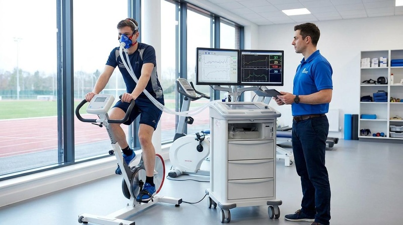
| Mechanism | Breath-by-breath gas exchange analysis ($\text{O}_2$ uptake, $\text{CO}_2$ output, ventilation) |
| Key Spec | $\dot{V}\text{O}_2\text{peak}$, $\dot{V}\text{E}/\\dot{V}\text{CO}_2$ slope, RER, Oxygen Pulse |
| Protocol | Incremental exercise (Ramp or Step) to volitional exhaustion (8–12 min) |
| Distance | Treadmill or Cycle Ergometer |
| FDA Class | Class II (Metabolic carts and gas analyzers) |
| Entry Cost | $150 – $500 per test clinical; $15,000+ for clinical systems |
Exercise stress testing is a vital clinical and physiological methodology for directly evaluating cardiorespiratory fitness, identifying the multi-system limitations of exercise tolerance, and predicting prognostic risk in cardiovascular and pulmonary conditions. Unlike static resting biomarkers, dynamic exercise testing measures functional capacity under metabolic stress, providing highly sensitive, reproducible, and objective insights into a patient’s physiologic reserve, diagnostic pathophysiology, and clinical prognosis.
¶ At a glance
Key points
- Does it work?: Yes. Cardiopulmonary exercise testing (CPET) and advanced exercise stress testing represent crucial clinical modalities for directly evaluating cardiorespiratory fitness, identifying the multi-system limitations of exercise tolerance, and predicting prognostic risk [1][2][3][4].
- Who needs it?: Indispensable for preoperative risk stratification before major high-risk surgeries (such as lung resections) [5], patients presenting with unexplained exertional dyspnea where resting diagnostics are inconclusive [2:1][3:1][4:1], heart failure patients requiring clinical phenotyping and risk assessment [1:1][6][7], and heart transplant recipients undergoing rehabilitation [8].
- Verdict: CPET and advanced exercise stress testing are the premier multi-system clinical probes. They replace subjective functional assessments with direct, objective physiological metrics, serving as a core pillar of modern clinical decision-making, patient selection, and exercise prescription [1:2][2:2][3:2][4:2].
¶ Evidence summary table (human outcomes)
| Outcome / Goal | Effect* | Consistency** | Evidence quality | Trials*** | Notes (population, duration, dose) |
|---|---|---|---|---|---|
| Preoperative Risk Stratification | High | High | Consensus Guidelines | Preoperative physiological assessment is recommended prior to major thoracic and high-risk surgical resections to evaluate functional capacity and stratify perioperative risk [5:1]. | |
| Functional Assessment in Heart Failure | High | High | Consensus Guidelines | Peak oxygen consumption () and ventilatory parameters are key tools used in clinical guidelines to stratify risk and evaluate functional impairment in patients with chronic heart failure [6:1][4:3][7:1]. | |
| Exercise Rehabilitation in Chronic Heart Failure | High | High | Clinical Trials | Structured aerobic exercise training is safe and well-tolerated in medically stable outpatients with heart failure with reduced ejection fraction (HFrEF) and improves health status [1:3][9]. | |
| Evaluation of Unexplained Dyspnea | High | High | Consensus Guidelines | In patients presenting with unexplained exertional dyspnea where resting diagnostic tests are inconclusive, cardiopulmonary exercise testing is recommended to differentiate between cardiac, pulmonary, and skeletal muscle limitations [2:3][4:4]. | |
| Detection of Pulmonary Hypertension in Systemic Sclerosis | High | Moderate | Cohort Studies | Cardiopulmonary exercise testing parameters, specifically peak oxygen uptake (), demonstrate high diagnostic accuracy for detecting pulmonary arterial hypertension in systemic sclerosis [10]. |
- *Effect: Compact renderer encoding represents direction, magnitude, and impact. Format: e="[dir][mag][impact]" where dir = u/d/e/q, mag = 0/1/2/3, and impact = p/n/x. (e.g., e="u3p" for large positive increase, e="d3p" for large positive decrease).
- **Consistency: Low (results conflict), Moderate (mixed but leaning one way), High (most trials agree)
- ***Trials: Level of evidence in the literature (e.g. Meta-analyses, Cohort studies, Consensus guidelines)
- REQUIRED: You MUST include a citation key (e.g.
[^1]) in the "Notes" column for every single row. If you claim a result, you must link the specific Meta-Analysis or Key RCT that proves it.
¶ Physiological Limitations of Heart Rate-Based Submaximal Estimation Models
While modern consumer fitness wearables offer a convenient means of longitudinal tracking, they rely on indirect heart rate-based submaximal estimation models rather than direct gas exchange analysis. Clinical guidelines and physiological consensus highlight several inherent limitations in these submaximal estimation models, which directly affect the accuracy of the fitness estimates produced by modern consumer devices:
- Mathematical and Linear Assumptions: Traditional heart rate-based estimations of cardiorespiratory fitness () assume a strict linear relationship between heart rate, oxygen consumption, and work rate [2:4][3:3][11][4:5]. Clinical guidelines demonstrate that this assumed linearity often breaks down, especially at high-intensity workloads or in clinical cohorts, leading to significant and unpredictable estimation errors [2:5][3:4][4:6][12].
- Autonomic and Environmental Confounders: Submaximal estimation models are highly sensitive to confounding physiological variables that alter the heart rate-workload relationship. For example, cardiac drift (the progressive, time-dependent increase in heart rate during prolonged, constant-load exercise due to thermoregulatory demands, mild dehydration, and peripheral blood pooling) artificially inflates heart rate relative to actual metabolic demand [2:6][3:5][11:1][4:7]. Similarly, factors such as ambient temperature, altitude, psychological stress, and sleep deprivation alter submaximal heart rate, causing estimation algorithms to incorrectly estimate aerobic capacity.
- Pharmacological Interference: In individuals taking chronotropic-altering medications (most notably beta-blockers), the physiological heart rate response to exercise is pharmacologically blunted [2:7][3:6][11:2]. Because standard submaximal heart rate estimation models are built on normal, unblocked sinus node responses, they cannot resolve these pharmacodynamic alterations, resulting in highly inaccurate and clinically misleading fitness estimates.
- Diagnostic Non-Equivalence: Heart rate-based submaximal models are entirely unable to capture breath-by-breath gas exchange dynamics. They cannot measure respiratory exchange ratio (RER), ventilatory efficiency ( slope), end-tidal gas fractions ( and ), or ventilatory thresholds [2:8][3:7][4:8]. Consequently, while helpful for general fitness tracking, they cannot serve as clinical diagnostic tools to identify underlying cardiovascular, pulmonary, or neuromuscular pathologies [2:9][3:8][4:9].
¶ Mechanism
¶ Wasserman's Gear Diagram Explanation
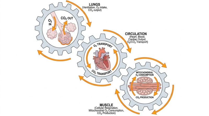
Karlman Wasserman conceptualized exercise physiology through three interlocking gears:
- Pulmonary Ventilation (Lungs): Regulates oxygen () intake and carbon dioxide () clearance via alveolar-capillary diffusion.
- Cardiovascular Circulation (Heart & Blood): Transports oxygenated blood to tissues, governed by cardiac output () and systemic blood flow.
- Cellular Respiration (Skeletal Muscle Mitochondria): Utilizes oxygen to generate ATP via oxidative phosphorylation, producing carbon dioxide.
During exercise testing, analyzing gas exchange allows clinicians to observe the integrity of these gears. In conditions like heart failure or pulmonary vascular diseases, the dysfunction of one gear (e.g., impaired cardiac output or pulmonary vascular resistance) impairs the entire chain, reducing peak exercise capacity and elevating ventilatory slopes [2:10][3:9][12:1][4:10].
¶ VO₂peak vs. VO₂max
- (maximal oxygen consumption, see VO₂ Max Training) represents the physiological ceiling of the cardiorespiratory system. It is strictly characterized by a plateau in oxygen consumption despite a further increase in workload [2:11][3:10][11:3][4:11].
- is the highest value of oxygen uptake attained during an incremental exercise test to volitional exhaustion, where a true plateau is not observed.
- In clinical settings and pathological cohorts, a true plateau is rarely achieved because patients are frequently limited by dyspnea, localized leg fatigue, or hemodynamic instability. Thus, is the primary metric reported in cardiovascular and respiratory medicine [1:4][6:2][4:12].
- When a true plateau in oxygen consumption is not achieved, measuring the respiratory exchange ratio (RER) provides an objective noninvasive indicator to help evaluate whether a patient has approached maximal physiologic effort [2:12][4:13].
¶ Respiratory Exchange Ratio (RER)
The Respiratory Exchange Ratio (RER) is the ratio of carbon dioxide production to oxygen consumption () measured at the mouth.
- Substrate Utilization: RER reflects the balance of metabolic substrate utilization and, during intense exercise, the buffering of lactic acid [2:13][3:11][11:4][4:14].
- Physiological Indicator: An increase in the respiratory exchange ratio (RER) is measured during clinical exercise testing as an objective indicator of gas exchange dynamics and physiological response [2:14][4:15].
¶ Oxygen Pulse (O₂ Pulse)
Oxygen Pulse is calculated as oxygen consumption divided by heart rate (), representing the volume of oxygen consumed per heartbeat. By rearranging the Fick Equation (), we find that:
Where is stroke volume and is the arteriovenous oxygen difference.
- Clinical Utility: Because reaches a predictable near-maximal extraction rate during heavy exercise, the trajectory of the pulse serves as a non-invasive surrogate for cardiac stroke volume dynamics [2:15][3:12][4:16].
- Ischemia and Dysfunction: In a healthy individual, the pulse rises progressively during incremental exercise and plateaus near peak capacity. An abrupt, premature flattening, a downward drop, or a chaotic fluctuation in the pulse is a highly sensitive clinical marker of myocardial ischemia, severe left ventricular dysfunction, valvular insufficiency, or an exercise-induced drop in stroke volume [2:16][3:13][4:17].
¶ Ventilatory Efficiency (VE/VCO₂ Slope)
The slope represents the efficiency of the respiratory system in clearing carbon dioxide. It is the slope of the linear relationship between minute ventilation () and carbon dioxide output () from the beginning of exercise throughout the physical effort [2:17][3:14][4:18].
- Pathophysiology: An elevated slope reflects ventilatory inefficiency, driven predominantly by ventilation-perfusion () mismatching, increased physiologic dead space, or hypersensitivity of peripheral and central chemoreceptors [2:18][3:15][4:19].
- Prognostic Power: The slope is one of the most powerful, load-independent prognostic markers in cardiovascular and respiratory medicine. In patients with chronic heart failure or pulmonary vascular diseases, an elevated slope is strongly predictive of cardiac hospitalization, transplant necessity, and mortality [2:19][3:16][4:20][13][14][15][16].
¶ Key Physiological and Hemodynamic Parameters
Cardiopulmonary exercise testing and advanced stress testing monitor a suite of physiological and hemodynamic parameters that provide deep insights into a patient’s resting and exertional physiology [2:20][3:17][4:21].
¶ Peak Oxygen Uptake (VO₂peak)
- Clinical Significance: represents the highest rate of oxygen consumption achieved during exercise and is the gold standard metric of cardiorespiratory fitness [2:21][3:18][4:22]. Relative to age- and sex-adjusted predicted normative values, an impaired peak oxygen consumption indicates functional capacity reduction [4:23].
- Preoperative Fitness Assessment: In candidates for lung cancer resection, clinical guidelines recommend a physiological assessment incorporating spirometry, diffusing capacity of carbon monoxide (), and exercise testing to stratify perioperative risk and guide surgical suitability [5:2]. According to the ACCP guidelines, a peak or predicted indicates high perioperative risk [5:3].
¶ Ventilatory Efficiency (VE/VCO₂ Slope)
- Clinical Significance: The slope measures the relationship between minute ventilation and carbon dioxide output, serving as an index of gas exchange efficiency [2:22][3:19][4:24].
- Pathophysiological Marker: An elevated slope reflects pulmonary vascular and ventilatory mismatching, which is highly prevalent in pulmonary vascular disease and chronic heart failure [2:23][3:20][4:25]. It is a powerful, load-independent predictor of death or the need for urgent heart transplantation [2:24][3:21][4:26][13:1][14:1][15:1][16:1].
¶ Exercise Pulmonary Hemodynamics
- Exercise Pulmonary Hemodynamics: Exercise pulmonary hypertension describes a condition of abnormal pulmonary hemodynamic response characterized by an excessive pulmonary arterial pressure increase in relation to flow during exercise, representing early pulmonary vascular disease, left heart disease, or lung disease [17].
- Hemodynamic Reserve in Pulmonary Vascular Disease: In early-stage pulmonary vascular disease, the pulmonary vasculature fails to accommodate the physiological increase in cardiac output during exercise. This results in an excessive increase in pulmonary arterial pressure relative to flow, which can be clinically unmasked during exercise right heart catheterization or stress echocardiography [17:1].
¶ Diastolic Stress Echocardiography (Left Ventricular Filling Pressures)
- Clinical Significance & Diastolic Impairment: Resting non-invasive echocardiographic parameters, such as the early mitral inflow to tissue velocity ratio (), demonstrate only modest or poor correlations with invasively measured left ventricular filling pressures and clinical outcomes in patients with heart failure with preserved ejection fraction (HFpEF) [18]. Because resting markers frequently fail to adequately reflect the hemodynamic burden experienced during exertion, dynamic exercise stress-testing models are clinically superior for capturing diastolic impairment, unmasking latent diastolic dysfunction, and documenting exertional elevations in left ventricular filling pressures [19][20][21].
¶ Testing Modalities and Protocols
The selection of the exercise modality and testing protocol is tailored to the patient's clinical presentation, functional baseline, and the specific diagnostic question being addressed [1:5][2:25][3:22][4:27].
¶ Exercise vs. Pharmacological Stress Testing
- Exercise Stress Testing: Active exercise on a treadmill or cycle ergometer represents the physiological gold standard, allowing for the direct evaluation of functional capacity, gas exchange, and multi-system exercise limitations [1:6][2:26][3:23][4:28].
- Pharmacological Stress Testing: When physical exercise is not feasible due to orthopedic, neurological, or other physical limitations, pharmacological stress testing may be utilized [11:5]. These protocols facilitate the assessment of myocardial perfusion or coronary flow reserve without requiring active physical exertion [11:6].
¶ The Role of Supine Bicycle Ergometry
- Diastolic Stress Echocardiography: Supine bicycle ergometry is highly preferred and recommended for evaluating patients with suspected heart failure with preserved ejection fraction (HFpEF) when resting diagnostics are equivocal [19:1][20:1][21:1].
- In-Test Imaging: Testing on a bicycle ergometer is highly preferred because it facilitates echocardiographic imaging during active exercise, providing a robust noninvasive method to diagnose HFpEF by identifying dynamic, exercise-induced elevations in left ventricular filling pressures and pulmonary pressures [19:2][4:29][20:2][21:2].
¶ Equipment Specifications and Calibration
To maintain quality control in clinical exercise laboratories, standard guidelines recommend structured calibration of treadmill, cycle ergometer, and metabolic systems to ensure accurate measurements of gas exchange and ventilation parameters [12:2].
- Calibration Requirements: Volume calibration is typically performed to verify flow sensor accuracy, while gas calibration utilizes high-precision calibration gas cylinders containing certified concentrations of oxygen and carbon dioxide to calibrate the relative gas analyzers [12:3].
- Mask Leaks and Fit: Mask selection is a critical determinant of measurement integrity. Undetected mask leaks falsely lower recorded minute ventilation (), , and , leading to an underestimated peak capacity and artificially skewed RER values.
- Ambient Conditions: Clinical gas analysis is highly sensitive to ambient temperature, humidity, and barometric pressure. Metabolic software processes respiratory variables (such as oxygen uptake and minute ventilation in BTPS) to provide reliable clinical data [2:27].
- Measurement Error Margin: Under optimal calibration and clinical execution, measurement error is kept to a minimum to ensure test reproducibility, but can increase significantly in the presence of uncorrected mask leaks, improper gas calibration, or uncompensated drift in laboratory temperature [2:28][12:4].
¶ Treadmill vs. Cycle Ergometer Protocols
Clinical CPET is performed utilizing either a treadmill or an electronically braked cycle ergometer, using either a Ramp protocol (where work rate increases continuously and smoothly) or a Step protocol (where work rate increases in discrete stages, e.g., 2-3 minute intervals) [2:29][3:24][4:30].
Ramp protocols are heavily preferred in clinical gas analysis because they facilitate a steady, linear rise in oxygen consumption and ventilation, permitting precise identification of the ventilatory anaerobic threshold (VAT) and respiratory compensation point (RCP) [2:30][3:25][4:31].
The selection between treadmill and cycle ergometer testing involves distinct biomechanical and physiological trade-offs:
| Feature / Param | Treadmill Testing | Cycle Ergometer Testing | Clinical Implications |
|---|---|---|---|
| Peak VO₂ Yield | Higher peak [2:31][3:26][11:7][4:32] | Lower peak due to smaller active muscle mass | Treadmill is preferred for maximal aerobic capacity profiling in athletes or general fitness. |
| Hemodynamic Monitoring | Technically challenging due to motion artifact | Significantly easier blood pressure monitoring and ECG tracing | Cycle ergometry is preferred when precise blood pressure monitoring or blood sampling (e.g., lactate) is required. |
| Local Muscle Fatigue | Less common; limited by overall cardiorespiratory capacity | Common; untrained patients are often limited by local quadriceps fatigue | Deconditioned or elderly patients may stop cycle testing early due to local leg fatigue before reaching cardiovascular peak. |
| Patient Safety & Joint Stress | Higher risk of balance loss or falls; weight-bearing | Safer, non-weight bearing; lower risk of falls or severe joint impact | Cycle is highly preferred for patients with severe orthopedic limitations, balance disorders, or advanced osteoarthritis [2:32][3:27][4:33]. |
¶ Indications, Contraindications, and Test Termination Criteria
¶ Clinical Indications
CPET is indicated for the following clinical and professional scenarios [2:33][3:28][4:34]:
- Unexplained Exertional Dyspnea: Discerning the relative contributions of cardiac, respiratory, hematologic, or deconditioning limitations when resting pulmonary and cardiac diagnostics are inconclusive [2:34][4:35].
- Heart Failure Prognosis and Stratification: Predicting risk of hospitalization, guiding listing for heart transplantation (where peak and ventilatory efficiency serve as primary clinical indicators [6:3][22][7:2]), and monitoring therapeutic responses in HFrEF and HFpEF [1:7][6:4][7:3].
- Preoperative Risk Stratification: Characterizing physical reserve before major, high-risk thoracic or abdominal surgeries (such as lung resection or radical cystectomy) to predict surgical risk [5:4].
- Evaluation of Unexplained Exercise Limitation: Characterizing dynamic metabolic disorders, mitochondrial dysfunction, or chronic fatigue syndromes.
- Sports Performance and Aerobic Profiling: Determining precise metabolic thresholds and peak oxygen consumption in elite athletes [11:8].
¶ Absolute Contraindications
CPET must NEVER be initiated in patients presenting with any of the following acute conditions [2:35][3:29][11:9]:
- Acute Myocardial Infarction (AMI) within the preceding 2 days
- Unstable Angina or acute coronary syndrome
- Uncontrolled Cardiac Arrhythmias causing symptoms or hemodynamic compromise
- Acute Myocarditis or Pericarditis
- Severe Symptomatic Aortic Stenosis
- Acute Pulmonary Embolism (PE) or pulmonary infarction
- Acute Deep Vein Thrombosis (DVT)
- Active Infective Endocarditis
- Acute Aortic Dissection
¶ Relative Contraindications
In patients with relative contraindications, testing may proceed only if the diagnostic value outweighs the hazard, under direct medical supervision [2:36][3:30][11:10]:
- Left Main Coronary Artery Stenosis (or its equivalent)
- Moderate Stenotic Valvular Heart Disease
- Severe Arterial Hypertension at Rest (Systolic Blood Pressure or Diastolic Blood Pressure )
- Tachyarrhythmias or Bradyarrhythmias with uncontrolled rates
- High-Degree Atrioventricular (AV) Block (Second-degree Mobitz Type II or Third-degree AV block)
- Hypertrophic Obstructive Cardiomyopathy (HOCM)
- Severe Mental or Physical Impairment preventing safe exercise performance
¶ Test Termination Criteria
To ensure clinical laboratory safety, CPET must be stopped immediately if any of the following safe-stopping criteria are observed [2:37][3:31][11:11]:
- ST-Segment Elevation in any lead without diagnostic Q waves [12:5][11:12].
- Progressive Drop in Systolic Blood Pressure from baseline despite an increase in workload, accompanied by objective signs of myocardial ischemia.
- Moderate-to-Severe Angina (Grade 3 or 4 on the standard angina scale).
- Central Nervous System Symptoms: Onset of ataxia, dizziness, confusion, or near-syncope.
- Signs of Poor Perfusion: Cyanosis, profound pallor, or cold, clammy skin.
- Technical Failure: Loss of continuous ECG recording or reliable blood pressure monitoring.
- Sustained Ventricular Tachycardia (VT) or high-grade ectopy (e.g., multifocal PVCs, runs of non-sustained VT).
- Patient's Request: Immediate volitional termination requested by the patient due to severe discomfort or exhaustion.
¶ Exercise Prescription and Rehabilitation
The clinical results of cardiopulmonary exercise testing are used to design safe, individualized exercise prescriptions, guiding therapeutic cardiac rehabilitation:
- Chronic Heart Failure: Standard clinical guidelines recommend that aerobic exercise training be considered for stable outpatients with heart failure to improve functional capacity and quality of life [1:8][6:5].
- Heart Transplantation Recipients: Prevention and rehabilitation programs after heart transplantation are recommended to be initiated early and continued post-transplant as multidisciplinary interventions to optimize physical capacity and long-term survival [8:1].
¶ Exercise Intensity and Training Zones
Aerobic exercise prescription is essential for optimizing improvements in exercise capacity while ensuring safety during cardiac rehabilitation [11:13]. Clinical standards support the use of exercise testing to define training intensity zones based on parameters such as peak heart rate, heart rate reserve, or ventilatory thresholds [11:14]. Rather than relying on rigid estimations, utilizing objective parameters from a graded exercise test allows clinicians to prescribe individualized, safe, and effective training zones (ranging from moderate to vigorous intensity) tailored to the patient’s functional capacity [11:15].
¶ Inaccuracy of Age-Predicted Formulas
Standard fitness programs and consumer wearables estimate training zones using heart rate maximum (HRmax) formulas, such as the classic equation.
This population-derived estimate possesses severe clinical limitations:
- High Individual Variance: Peer-reviewed studies demonstrate that the standard deviation for the equation carries a large and clinically significant individual variance [3:32][11:16]. This means that for a 40-year-old with an estimated HRmax of , their actual biological HRmax can differ substantially from this value.
- Risk of Inappropriate Stimulus: Utilizing an inaccurate estimated maximum to define training intensity can lead to patients unconsciously training in a higher metabolic intensity zone, causing chronic overtraining, elevated cortisol, and blunted mitochondrial adaptations.
- Clinical Safety Risks: In cardiovascular cohorts, using age-predicted formulas to prescribe exercise intensity can lead to prescribing target heart rates that exceed the patient's ischemic threshold or provoke unstable arrhythmias, creating severe clinical safety hazards [2:38][3:33][11:17].
¶ Clinical Interpretation by Trained Clinicians
The clinical interpretation of cardiopulmonary exercise testing requires a sophisticated, comprehensive integration of multiple physiological systems. A raw CPET report cannot be interpreted by a single metric (like peak ) in isolation, nor can it be self-interpreted by patients or estimated using consumer-facing proxies.
A valid CPET interpretation requires [2:39][3:34][4:36]:
- Multi-System Integration: The interpreter must integrate cardiovascular function (ECG, blood pressure response, oxygen pulse), respiratory function (ventilatory reserve, breathing reserve, gas exchange dynamics, ), hematologic capacity (oxygen-carrying capacity), and neuromuscular/skeletal muscle metabolic capacity (work rate, VAT, local muscle fatigue) [2:40][3:35][4:37].
- Clinical Specialization: Interpretation of clinical CPET diagnostic cascades should be performed by qualified healthcare professionals (such as trained cardiologists, pulmonologists, or clinical exercise specialists) who possess specific training in exercise physiology and gas exchange analysis [2:41][3:36].
- Clinical Warning: Self-interpretation of raw metabolic cart outputs or relying on consumer wearable proxies is strongly discouraged. It carries a high risk of misinterpreting normal physiological responses as pathological, or conversely, missing critical early markers of cardiac ischemia, valvular disease, or pulmonary vascular disease [2:42][3:37][4:38].
¶ Stepwise Interpretation Algorithm
Clinical interpretation of exercise testing relies on a structured, multi-system pathophysiological algorithm rather than analyzing metrics in isolation. This system-level assessment differentiates between respiratory, cardiac, circulatory, and skeletal muscle exercise limitations [1:9][6:6][2:43][3:38][4:39].
¶ Stepwise Diagnostic Cascade
A clinical interpreter follows a stepwise diagnostic cascade to decode exercise capacity [2:44][3:39][4:40]:
- Evaluate Peak Effort: Verify RER and ventilatory parameters to ensure a valid effort is achieved [2:45][3:40][4:41].
- Assess Aerobic Capacity: Compare peak to predicted reference normative values. A significant reduction relative to predicted values indicates compromised aerobic capacity and functional impairment [2:46][3:41][4:42].
- Evaluate Diastolic and Hemodynamic Function: Compare resting diastolic function metrics. Resting measurements of diastolic parameters correlate poorly or modestly with invasive hemodynamics and may fail to reflect exertional symptoms [18:1]. When resting diagnostics are inconclusive, dynamic stress-testing models are superior for capturing diastolic impairment, unmasking latent diastolic dysfunction, and identifying dynamic, exercise-induced elevations in left ventricular filling pressures [19:3][20:3][21:3].
- Inspect Ventilatory Efficiency: Examine the slope. An elevated slope suggests ventilatory inefficiency, driven by ventilation-perfusion () mismatching, highly characteristic of heart failure or pulmonary vascular diseases [2:47][3:42][4:43][13:2].
¶ Evidence
¶ Clinical Outcomes and Prognostic Utility
The peer-reviewed clinical literature demonstrates that exercise testing provides unmatched prognostic and risk-prediction data across multiple patient populations:
- Preoperative Risk Stratification:
Preoperative functional status is a major predictor of surgical outcomes. For patients undergoing lung resection, cardiopulmonary exercise testing is indicated to evaluate aerobic capacity and stratify perioperative risk when lung function is impaired [5:5]. The 2013 American College of Chest Physicians (ACCP) guidelines recommend utilizing peak to define surgical risk [5:6]. A peak or predicted represents a high risk of mortality and complications for major anatomic resections [5:7]. - Heart Failure Management:
CPET-derived parameters are paramount for predicting outcomes in cardiovascular disease. In patients with chronic heart failure, depressed peak and ventilatory efficiency are critical markers used to evaluate functional impairment, predict clinical outcomes, and guide listings for heart transplantation [6:7][4:44][7:4]. In clinical practice, parameters of peak and percent-predicted peak are standard parameters generally used as objective functional thresholds to guide selection for advanced heart failure therapies [6:8][4:45][7:5]. Under these general clinical standards, the prognostic evaluation of exercise capacity serves to stratify risk and guide transplant consideration [6:9][4:46][22:1][7:6]. - Pulmonary Vascular Disease:
In patients with systemic sclerosis, pulmonary hypertension is a heterogeneous and progressive condition that represents a major cause of morbidity and mortality [13:3][23]. Cardiopulmonary exercise testing parameters, particularly peak oxygen uptake, show high diagnostic accuracy for identifying associated pulmonary hypertension and characterizing functional impairment in patients with systemic sclerosis [10:1]. Clinical studies demonstrate that CPET parameters correlate significantly with invasive pulmonary hemodynamics; specifically, a peak oxygen uptake () of shows high sensitivity and specificity for detecting systemic sclerosis-associated pulmonary arterial hypertension, whereas a peak can reliably exclude it [10:2]. Furthermore, longitudinal studies of systemic sclerosis patients without resting pulmonary hypertension show that abnormal increases in exercise pulmonary arterial pressure and reductions in peak over time serve as indicators of early pulmonary vascular disease progression [24]. While CPET is highly beneficial in non-invasively characterizing functional impairment, clinical reviews emphasize that systemic sclerosis-associated pulmonary hypertension is highly complex, and the optimal diagnostic thresholds as well as the exact role of exercise in identifying early disease require further clinical elucidation [23:1]. - Exercise Training and Rehabilitation:
In medically stable outpatients with heart failure and reduced ejection fraction, a structured program of aerobic exercise training (consisting of supervised sessions followed by home-based training) was shown in the HF-ACTION trial to be safe, with other adverse events being similar to usual care [1:10], and resulted in modest improvements in self-reported health status [9:1]. Furthermore, a clinical consensus statement emphasizes the importance of prevention and rehabilitation programs after heart transplantation (HTx), which should be specifically tailored, multidisciplinary in nature, initiated early after surgery, and continued throughout the post-transplant journey to address modifiable and non-modifiable factors to improve physical capacity, quality of life, and survival [8:2].
¶ Safety
¶ Safety, Screening, and Supervision Standards
Clinical exercise laboratories must operate under strict, standardized safety and quality-control protocols to protect patients while achieving a valid maximal diagnostic effort [12:6][11:18].
- Supervision Standards: Structured exercise training and clinical testing programs have an exceptionally high safety profile. Large-scale trials, such as the HF-ACTION study, demonstrate that structured exercise training is highly safe and well-tolerated in medically stable outpatients with chronic heart failure [1:11].
- Pre-Test Screening: Candidates with chronic conditions must be clinically stable before undergoing exercise testing or participating in supervised exercise programs [1:12][2:48][3:43][4:47]. Clinical exercise laboratories must establish clear guidelines regarding personnel qualifications and emergency preparation to manage any potential acute adverse events during testing [12:7].
¶ FAQ
- What is the difference between VO₂max and VO₂peak?:
VO₂max represents a true physiological ceiling and is defined by a plateau in oxygen uptake despite an increase in exercise intensity. VO₂peak is simply the highest value of oxygen uptake achieved during an exercise test to volitional exhaustion, without meeting the strict plateau criteria. In clinical testing, VO₂peak is the standard metric used [1:13][6:10][2:49][3:44][4:48]. - Is exercise training safe in heart failure?:
Yes, clinical research confirms that structured aerobic exercise training is safe and well-tolerated in medically stable outpatients with heart failure. Large-scale trials show that exercise training does not increase adverse clinical events and is associated with significant long-term clinical benefits when combined with optimal medical therapy [1:14][2:50][3:45][4:49]. - Why is stress testing used in heart failure with preserved ejection fraction (HFpEF)?:
Many patients with HFpEF have normal resting diagnostics but experience severe dyspnea during exertion. Because resting echocardiographic markers correlate poorly with invasive hemodynamics, dynamic stress-testing models are superior for capturing diastolic impairment. When resting assessments are inconclusive, dynamic testing under exercise stress is valuable to unmask latent diastolic dysfunction and demonstrate elevated filling pressures [19:4][20:4][21:4]. - How does CPET help predict risk in pulmonary vascular disease?:
In patients with systemic sclerosis or other high-risk cohorts, CPET-derived parameters serve as valuable non-invasive diagnostic and risk-prediction tools. Specifically, peak oxygen consumption () and ventilatory efficiency are strongly correlated with invasive hemodynamics and can identify early-stage pulmonary vascular disease and functional impairment [23:2][10:3][24:1].
¶ References
O'Connor CM, Whellan DJ, Lee KL, et al. Efficacy and safety of exercise training in patients with chronic heart failure: HF-ACTION randomized controlled trial. JAMA. 2009;301(14):1439-1450. https://pubmed.ncbi.nlm.nih.gov/19351941/ ↩︎ ↩︎ ↩︎ ↩︎ ↩︎ ↩︎ ↩︎ ↩︎ ↩︎ ↩︎ ↩︎ ↩︎ ↩︎ ↩︎ ↩︎
Ross RM. ATS/ACCP Statement on cardiopulmonary exercise testing. American Journal of Respiratory and Critical Care Medicine. 2003;167(10):1451. https://pubmed.ncbi.nlm.nih.gov/12738602/ ↩︎ ↩︎ ↩︎ ↩︎ ↩︎ ↩︎ ↩︎ ↩︎ ↩︎ ↩︎ ↩︎ ↩︎ ↩︎ ↩︎ ↩︎ ↩︎ ↩︎ ↩︎ ↩︎ ↩︎ ↩︎ ↩︎ ↩︎ ↩︎ ↩︎ ↩︎ ↩︎ ↩︎ ↩︎ ↩︎ ↩︎ ↩︎ ↩︎ ↩︎ ↩︎ ↩︎ ↩︎ ↩︎ ↩︎ ↩︎ ↩︎ ↩︎ ↩︎ ↩︎ ↩︎ ↩︎ ↩︎ ↩︎ ↩︎ ↩︎ ↩︎
Balady GJ, Arena R, Sietsema K, et al. Clinician's Guide to cardiopulmonary exercise testing in adults: a scientific statement from the American Heart Association. Circulation. 2010;122(2):191-225. https://pubmed.ncbi.nlm.nih.gov/20585013/ ↩︎ ↩︎ ↩︎ ↩︎ ↩︎ ↩︎ ↩︎ ↩︎ ↩︎ ↩︎ ↩︎ ↩︎ ↩︎ ↩︎ ↩︎ ↩︎ ↩︎ ↩︎ ↩︎ ↩︎ ↩︎ ↩︎ ↩︎ ↩︎ ↩︎ ↩︎ ↩︎ ↩︎ ↩︎ ↩︎ ↩︎ ↩︎ ↩︎ ↩︎ ↩︎ ↩︎ ↩︎ ↩︎ ↩︎ ↩︎ ↩︎ ↩︎ ↩︎ ↩︎ ↩︎ ↩︎
Arena R, Myers J, Williams MA, et al. Assessment of functional capacity in clinical and research settings: a scientific statement from the American Heart Association Committee on Exercise, Rehabilitation, and Prevention of the Council on Clinical Cardiology and the Council on Cardiovascular Nursing. Circulation. 2007;116(3):329-343. https://pubmed.ncbi.nlm.nih.gov/17576872/ ↩︎ ↩︎ ↩︎ ↩︎ ↩︎ ↩︎ ↩︎ ↩︎ ↩︎ ↩︎ ↩︎ ↩︎ ↩︎ ↩︎ ↩︎ ↩︎ ↩︎ ↩︎ ↩︎ ↩︎ ↩︎ ↩︎ ↩︎ ↩︎ ↩︎ ↩︎ ↩︎ ↩︎ ↩︎ ↩︎ ↩︎ ↩︎ ↩︎ ↩︎ ↩︎ ↩︎ ↩︎ ↩︎ ↩︎ ↩︎ ↩︎ ↩︎ ↩︎ ↩︎ ↩︎ ↩︎ ↩︎ ↩︎ ↩︎ ↩︎
Brunelli A, Kim AW, Berger KI, et al. Physiologic evaluation of the patient with lung cancer being considered for resectional surgery: Diagnosis and management of lung cancer, 3rd ed: American College of Chest Physicians evidence-based clinical practice guidelines. Chest. 2013;143(5 Suppl):e166S-e190S. https://pubmed.ncbi.nlm.nih.gov/23649437/ ↩︎ ↩︎ ↩︎ ↩︎ ↩︎ ↩︎ ↩︎ ↩︎
McMurray JJ, Adamopoulos S, Anker SD, et al. ESC Guidelines for the diagnosis and treatment of acute and chronic heart failure 2012: The Task Force for the Diagnosis and Treatment of Acute and Chronic Heart Failure 2012 of the European Society of Cardiology. Developed in collaboration with the Heart Failure Association (HFA) of the ESC. European Heart Journal. 2012;33(14):1787-1847. https://pubmed.ncbi.nlm.nih.gov/22611136/ ↩︎ ↩︎ ↩︎ ↩︎ ↩︎ ↩︎ ↩︎ ↩︎ ↩︎ ↩︎ ↩︎
Brawner CA, Lazar MH. Cardiopulmonary exercise testing criteria for advanced therapies in patients with heart failure. Heart Failure Reviews. 2023;28(6):1233-1244. https://pubmed.ncbi.nlm.nih.gov/37644366/ ↩︎ ↩︎ ↩︎ ↩︎ ↩︎ ↩︎ ↩︎
Simonenko M, Hansen D, Niebauer J, et al. Prevention and rehabilitation after heart transplantation: A clinical consensus statement of the European Association of Preventive Cardiology, Heart Failure Association of the ESC, and the European Cardio Thoracic Transplant Association, a section of ESOT. European Journal of Preventive Cardiology. 2024;31(14):1687-1702. https://pubmed.ncbi.nlm.nih.gov/38894688/ ↩︎ ↩︎ ↩︎
Flynn KE, Piña IL, Whellan DJ, et al. Effects of exercise training on health status in patients with chronic heart failure: HF-ACTION randomized controlled trial. JAMA. 2009;301(14):1451-1459. https://pubmed.ncbi.nlm.nih.gov/19351942/ ↩︎ ↩︎
Dumitrescu D, Nagel C, Kovacs G, et al. Cardiopulmonary exercise testing for detecting pulmonary arterial hypertension in systemic sclerosis. Heart. 2017;103(17):1288-1294. https://pubmed.ncbi.nlm.nih.gov/28062514/ ↩︎ ↩︎ ↩︎ ↩︎
Fletcher GF, Ades PA, Kligfield P, et al. Exercise standards for testing and training: a scientific statement from the American Heart Association. Circulation. 2013;128(8):873-934. https://pubmed.ncbi.nlm.nih.gov/23877260/ ↩︎ ↩︎ ↩︎ ↩︎ ↩︎ ↩︎ ↩︎ ↩︎ ↩︎ ↩︎ ↩︎ ↩︎ ↩︎ ↩︎ ↩︎ ↩︎ ↩︎ ↩︎ ↩︎
Myers J, Arena R, Franklin B, et al. Recommendations for clinical exercise laboratories: a scientific statement from the American Heart Association. Circulation. 2009;119(24):3144-3162. https://pubmed.ncbi.nlm.nih.gov/19487589/ ↩︎ ↩︎ ↩︎ ↩︎ ↩︎ ↩︎ ↩︎ ↩︎
Barbagelata L, Masson W, Bluro I, et al. Prognostic role of cardiopulmonary exercise testing in pulmonary hypertension: a systematic review and meta-analysis. Advances in Respiratory Medicine. 2022;90(2):87-94. https://pubmed.ncbi.nlm.nih.gov/35199845/ ↩︎ ↩︎ ↩︎ ↩︎
Cantone A, Serenelli M, Sanguettoli F, et al. Cardiopulmonary exercise testing predicts prognosis in amyloid cardiomyopathy: a systematic review and meta-analysis. ESC Heart Failure. 2023;10(4):2189-2199. https://pubmed.ncbi.nlm.nih.gov/37264762/ ↩︎ ↩︎
Baccelli A, Rinaldo RF, Haji G, et al. Prognostic value of cardiopulmonary exercise testing in pulmonary arterial hypertension. European Respiratory Journal. 2025;66(2):40210410. https://pubmed.ncbi.nlm.nih.gov/40210410/ ↩︎ ↩︎
Weatherald J, Farina S, Bruno N, et al. Cardiopulmonary Exercise Testing in Pulmonary Hypertension. Annals of the American Thoracic Society. 2017;14(Supplement 1):S84-S92. https://pubmed.ncbi.nlm.nih.gov/28375670/ ↩︎ ↩︎
Kovacs G, Herve P, Barbera JA, et al. An official European Respiratory Society statement: pulmonary haemodynamics during exercise. European Respiratory Journal. 2017;50(5):1700578. https://pubmed.ncbi.nlm.nih.gov/29167297/ ↩︎ ↩︎
Nauta JF, Hummel YM, van der Meer P, et al. Correlation with invasive left ventricular filling pressures and prognostic relevance of the echocardiographic diastolic parameters used in the 2016 ESC heart failure guidelines and in the 2016 ASE/EACVI recommendations: a systematic review in patients with heart failure with preserved ejection fraction. European Journal of Heart Failure. 2018;20(9):1303-1311. https://pubmed.ncbi.nlm.nih.gov/29877602/ ↩︎ ↩︎
Shim CY. Stress Testing in Heart Failure with Preserved Ejection Fraction. Heart Failure Clinics. 2021;17(3):405-414. https://pubmed.ncbi.nlm.nih.gov/34051975/ ↩︎ ↩︎ ↩︎ ↩︎ ↩︎
Yuasa N, Harada T, Kagami K, et al. The roles of exercise stress echocardiography for the evaluation of heart failure with preserved ejection fraction in the heart failure pandemic era. Journal of Medical Ultrasonics. 2024;51(3):389-401. https://pubmed.ncbi.nlm.nih.gov/38926301/ ↩︎ ↩︎ ↩︎ ↩︎ ↩︎
Guazzi M, Wilhelm M, Halle M, et al. Exercise testing in heart failure with preserved ejection fraction: an appraisal through diagnosis, pathophysiology and therapy - A clinical consensus statement of the Heart Failure Association and European Association of Preventive Cardiology of the European Society of Cardiology. European Journal of Heart Failure. 2022;24(8):1327-1345. https://pubmed.ncbi.nlm.nih.gov/35775383/ ↩︎ ↩︎ ↩︎ ↩︎ ↩︎
Felker GM, Whellan D, Kraus WE, et al. N-terminal pro-brain natriuretic peptide and exercise capacity in chronic heart failure: data from the Heart Failure and a Controlled Trial Investigating Outcomes of Exercise Training (HF-ACTION) study. American Heart Journal. 2009;158(4) Suppl:S144-S152. https://pubmed.ncbi.nlm.nih.gov/19782787/ ↩︎ ↩︎
Haque A, Kiely DG, Kovacs G, et al. Pulmonary hypertension phenotypes in patients with systemic sclerosis. European Respiratory Review. 2021;30(161):210047. https://pubmed.ncbi.nlm.nih.gov/34407977/ ↩︎ ↩︎ ↩︎
Kovacs G, Avian A, Wutte N, et al. Changes in pulmonary exercise haemodynamics in scleroderma: a 4-year prospective study. European Respiratory Journal. 2017;50(1):1601959. https://pubmed.ncbi.nlm.nih.gov/28705939/ ↩︎ ↩︎