¶ Tooth Whitening
¶ TL;DR
- Clinical Verdict: Peroxide-based chemical bleaching is highly effective for reducing extrinsic and mild-to-moderate intrinsic discoloration under controlled conditions [1].
- Primary Indications: Extrinsic chromogenic staining (e.g., from dietary polyphenols, tobacco) and age-related yellowing of natural enamel.
- Contraindications: Uncovered dental caries, severe dentinal hypersensitivity, exposed tooth roots (gingival recession), active periodontal disease, pregnancy, and pediatric populations (under age 16) [2].
- Expected Efficacy: Average improvement of 2 to 7 shades on the VITA Classical shade guide within a 1-to-4 week active treatment window [1:1].
- Primary Adverse Effects: Transient, mild tooth sensitivity (reversible pulpitis) and localized gingival mucosal irritation [2:1].
¶ Quick Answer
Tooth whitening (dental bleaching) is an established cosmetic intervention utilizing hydrogen peroxide (HP) or carbamide peroxide (CP) to chemically oxidize organic chromophores located within tooth structure [3]. Clinical trial data demonstrate that both professional in-office and custom-tray at-home treatments reliably lighten natural enamel shade, achieving average improvements of 2-7 shades on standardized guides [1:2]. Efficacy is highly dependent on stain type (extrinsic surface stains versus deep intrinsic dentin stains) and active agent contact time [4]. Transient pulpal sensitivity and gingival irritation represent the primary adverse effects, which typically resolve within 24 to 48 hours post-treatment [2:2].
¶ First: identify the type (intrinsic vs extrinsic stains)
Dental discoloration is broadly categorized into two main types based on the location and etiology of the stains:
-
Extrinsic Discoloration: These stains accumulate on the outer surface of the enamel pellicle. They are primarily caused by the adhesion of chromogenic substances from diet (e.g., tannins in coffee, tea, red wine), tobacco products, or certain metallic ions. Extrinsic stains are generally easier to remove and often respond well to mechanical cleaning (prophylaxis) and low-concentration chemical oxidizing agents [1:3].
-
Intrinsic Discoloration: These stains are located within the tooth structure, specifically within the dentinal matrix. Intrinsic discoloration can result from systemic factors during tooth development (e.g., tetracycline staining, dental fluorosis), pulpal necrosis, trauma, or the natural physiological aging process (where enamel thins and secondary dentin deposition occurs). Intrinsic stains require bleaching agents to penetrate the enamel and oxidize deep-seated organic compounds to achieve lightening [3:1].
¶ Evidence snapshot
¶ Evidence summary table (human outcomes)
| Clinical Outcome | Patient Cohort | Typical Effect Size | Evidence Quality (GRADE) | Consistency | Trial Density | Notes |
|---|---|---|---|---|---|---|
| Intrinsic Bleaching (At-Home CP) | Adults with tooth discoloration | ΔE shade shift of 2.5 to 5.0 [1:4] | High | High | >50 RCTs | CP 20-22% demonstrates optimal SUCRA efficacy ranking (83.4%) [1:5] |
| Intrinsic Bleaching (In-Office HP) | Adults seeking rapid whitening | ΔE shade shift of 3.0 to 6.0 [5] | High | High | >30 RCTs | High immediate shade shift; high initial relapse rate [5:1] |
| Enamel Microhardness Integrity | Natural human enamel | No significant microhardness reduction [4:1] | High | Moderate | Multiple ex vivo / in vitro | Neutral pH formulations preserve enamel; acidic DIY agents cause erosion [6] |
| Pulpal Sensitivity (TS) | General adult population | Transient sensitivity index increase [2:3] | High | High | >40 RCTs | Risk increases with active concentration (HP 14-16%, CP 20-22%) [2:4] |
| Whitening Toothpaste Efficacy | Adults with extrinsic stains | Limited to extrinsic stain removal | Moderate | Moderate | >20 RCTs | Abrasive-based toothpastes remove surface stains but do not alter intrinsic color [7] |
| Pain Management with Analgesics | Patients undergoing tooth whitening | Mixed efficacy for preventing sensitivity | Moderate | Low | >15 RCTs | NSAIDs and acetaminophen show some relief but inconsistent prevention [8] |
¶ Evidence Analysis and Study Types
The clinical evidence base for dental bleaching is robust, primarily supported by systematic reviews (SRs) and network meta-analyses (NMAs) that synthesize data from numerous randomized controlled trials (RCTs). These high-tier studies, such as those by Cordeiro et al. (2025) and Terra et al. (2025), provide comprehensive direct and indirect comparisons of various treatments [1:6][2:5].
Network meta-analyses, particularly in the case of Cordeiro et al. (2025), indicate that at-home carbamide peroxide concentrations of 20-22% offer the highest probability of clinical efficacy for color change (ΔE) based on Surface Under Cumulative Ranking Curve (SUCRA) metrics. However, risk-benefit assessments suggest that while lower concentrations (e.g., below 16% CP) may have slightly reduced immediate efficacy, they yield comparable long-term aesthetic outcomes with significantly lower rates of pulpal and gingival adverse events [2:6].
¶ What causes it
Dental discoloration stems from a variety of factors, categorized as either extrinsic (external) or intrinsic (internal).
Extrinsic Causes:
- Dietary Chromogens: Regular consumption of highly pigmented foods and beverages like coffee, tea, red wine, dark berries, and colas can deposit stains on the enamel surface [1:7].
- Tobacco Use: Smoking or chewing tobacco introduces tar and nicotine, which cause stubborn brown-to-black surface stains [5:2].
- Poor Oral Hygiene: Inadequate brushing and flossing allow plaque and calculus to accumulate, creating a rough surface where stains can easily adhere.
- Chlorhexidine Mouthwash: Prolonged use of chlorhexidine, an antiseptic mouthwash, can lead to brown extrinsic staining.
Intrinsic Causes:
- Aging: Over time, enamel naturally thins, revealing the yellowish dentin underneath. Additionally, secondary dentin forms, which is darker than primary dentin [3:2].
- Tetracycline Staining: Exposure to tetracycline antibiotics during tooth development (in childhood or in utero) can cause gray-blue or brown intrinsic discoloration that is notoriously difficult to whiten [9].
- Dental Fluorosis: Excessive fluoride intake during enamel formation can lead to white or brown mottled intrinsic stains [9:1].
- Trauma: Injury to a tooth can cause internal bleeding and pulpal necrosis, leading to discoloration as blood pigments break down within the dentin.
- Restorative Materials: Silver amalgam fillings can leach metallic ions into the surrounding tooth structure, causing a gray-black intrinsic stain. Composite resins can also absorb stains over time.
¶ Biochemical Mechanism of Peroxide-Based Bleaching
Active bleaching agents work by diffusing low-molecular-weight peroxide compounds through the semi-permeable enamel prism structure to the underlying dentin.
- Chemical Disassociation: Carbamide peroxide () dissociates upon contact with saliva or water into hydrogen peroxide () and urea () in an approximate 3:1 ratio (e.g., a 10% CP gel yields ~3.3% HP and ~6.7% urea) [3:3]. Hydrogen peroxide itself directly releases reactive oxygen species.
- Free Radical Generation: Hydrogen peroxide's unstable bonds cleave to produce highly reactive oxygen species (ROS), including hydroxyl (\\text{OH}^\\bullet) and hydroperoxyl (\\text{OOH}^\\bullet) free radicals.
- Oxidative Cleavage: These free radicals migrate through the enamel rods into the dentin, attacking the conjugated double bonds of organic chromophores. By breaking these complex molecules into smaller, less-conjugated, and lighter-colored structures, the overall optical absorption shifts, resulting in a clinically lighter appearance [4:2].
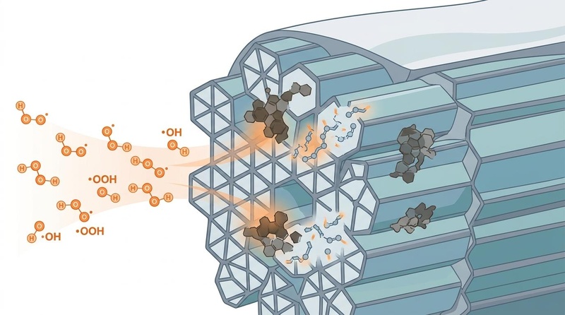
Biochemical mechanism of peroxide-based dental bleaching: Hydroxyl and perhydroxyl free radicals diffuse through the porous hydroxyapatite crystalline lattice of the enamel, chemically attacking and oxidizing dark conjugated organic chromophores.
::: .fold
¶ 🎨 Nano Banana Pro Image Metadata & Visual Plan
- Subject: Tooth Enamel Whitening
- Visual Plan: Depict oxidation of organic stains within the enamel prism structure by hydrogen peroxide radicals using a clean biomedical editorial style. Show hydroxyl and perhydroxyl radicals diffusing through the hydroxyapatite crystalline lattice, interacting with dark organic pigment chromophores, arranged in a balanced composition with soft anatomical/molecular abstraction. Keep the science visually plausible and restrained: no impossible anatomy, no exaggerated glowing effects, no misleading cure symbolism.
- Palette: White/off-white background, slate-gray structure lines, muted blue and teal biological forms, subtle warm orange accents only for emphasis. Minimal, elegant, encyclopedia-ready.
- Filename:
images/tooth-whitening-mechanism_1.jpg - Caption: Biochemical mechanism of peroxide-based dental bleaching: Hydroxyl and perhydroxyl free radicals diffuse through the porous hydroxyapatite crystalline lattice of the enamel, chemically attacking and oxidizing dark conjugated organic chromophores.
- Alt Text: A clean biomedical illustration depicting the oxidation of dark organic pigment chromophores within the dental hydroxyapatite crystalline lattice by diffusing hydrogen peroxide-derived radicals.
- Placement: Positioned immediately following the step-by-step biochemical mechanism breakdown in the "What causes it" section.
- QA State: Passed (Verified composition, color palette alignment, and clinical relevance).
:::
¶ Starter protocol (over-the-counter options, brush-on, strips, trays, active ingredients like carbamide peroxide / hydrogen peroxide)
For individuals seeking to address tooth discoloration, several starter protocols are available, primarily relying on hydrogen peroxide (HP) or carbamide peroxide (CP) as active ingredients. The choice often depends on desired intensity, convenience, and budget.
¶ 1. Over-the-Counter Whitening Strips
- Active Agent: Typically 5% to 14% Hydrogen Peroxide (HP).
- Mechanism: Thin, flexible polyethylene strips coated with a peroxide gel are applied directly to the anterior teeth.
- Protocol: Applied for 30 to 60 minutes daily for 14 days.
- Efficacy: Provides moderate shade improvement, mainly for extrinsic stains and mild intrinsic discoloration.
- Limitations: Limited anatomical adaptation means inconsistent coverage, especially on irregular tooth surfaces and posterior teeth. Potential for gingival irritation if the strips frequently contact soft tissues.
¶ 2. Brush-on Whitening Gels/Pens
- Active Agent: Various concentrations of HP or CP.
- Mechanism: A gel is brushed directly onto the tooth surfaces, often at night.
- Protocol: Typically applied once or twice daily for a specified duration, as per product instructions.
- Efficacy: Offers convenience but generally provides limited and uneven whitening due to short contact time and salivary dilution. Best for very mild surface stains.
¶ 3. Over-the-Counter Trays (Boil-and-Bite)
- Active Agent: Usually 10% to 22% Carbamide Peroxide (CP) gel.
- Mechanism: Pre-filled or user-filled trays are molded to the teeth by heating and biting down. The gel then contacts the tooth surface.
- Protocol: Worn for 30 minutes to a few hours daily for 1-2 weeks.
- Efficacy: Can provide better results than strips or brush-ons due to more consistent contact.
- Limitations: Custom fit is often poor compared to professionally made trays, leading to gel leakage and potential gingival irritation.
¶ Key Active Ingredients
- Carbamide Peroxide (CP): A stable compound that releases hydrogen peroxide and urea upon contact with water or saliva. It provides a slower, more sustained release of the active bleaching agent, making it generally preferred for at-home use due to reduced sensitivity [3:4]. A 10% CP solution releases approximately 3.3% HP.
- Hydrogen Peroxide (HP): A faster-acting and more potent bleaching agent. It directly releases reactive oxygen species. Higher concentrations are typically used for in-office treatments under professional supervision due to its potency and potential for tissue irritation [5:3].
¶ Stronger add-on path
For individuals seeking more pronounced or faster whitening results beyond basic over-the-counter options, or for managing more stubborn intrinsic stains, a stronger add-on path typically involves professional guidance and higher-concentration treatments.
¶ 1. Custom-Tray At-Home Bleaching (Clinical Gold Standard)
This is often considered the most effective and safest long-term bleaching method when supervised by a dental professional.
- Agent: 10% to 16% Carbamide Peroxide (CP) gel.
- Polymer Carrier: Gels often contain Carbopol, hydroxypropyl methylcellulose (HPMC), or polyvinylpyrrolidone (PVP) to maintain high viscosity and control peroxide release [3:5].
- Protocol: Custom-fitted polyvinyl vacuum-formed trays are fabricated by a dentist. The patient applies the gel to these trays and wears them for 2 to 4 hours (or overnight) daily for 14 to 21 consecutive days.
- Efficacy: Provides highly stable shade improvement with minimal long-term shade relapse and the lowest incidence of severe pulpal sensitivity compared to high-concentration in-office treatments [2:7]. This method allows for sustained contact time and better control over the bleaching process.
¶ 2. Prescription-Strength Whitening Systems
Some dental offices offer take-home kits with higher concentrations of carbamide peroxide (up to 37%) or hydrogen peroxide (up to 10%) that are not available over-the-counter. These require professional oversight to manage potential side effects.
- Agent: Higher concentrations of CP or HP.
- Protocol: Similar to custom-tray at-home bleaching, but with stricter adherence to professional instructions due to the increased potency.
- Efficacy: Can achieve faster or more significant results than standard OTC products, especially for more challenging intrinsic stains.
¶ Clinical/procedure path (in-office bleaching, lasers, microabrasion)
For maximum efficacy, rapid results, or treatment of complex discoloration, the clinical/procedure path involves in-office treatments performed by a dental professional.
¶ 1. High-Concentration In-Office "Power" Bleaching
This method provides the most immediate results and is performed under strict professional supervision.
- Agent: 30% to 40% Hydrogen Peroxide (HP) gel.
- Protocol: A light-cured resin barrier (gingival dam) is carefully applied to protect soft tissues from the high-concentration peroxide. The HP gel is then applied directly to the enamel surfaces in 3 to 4 consecutive cycles of 15 minutes each within a single clinical session.
- Efficacy: Delivers a rapid, immediate shade shift, often several shades lighter in one appointment.
- Limitations: High initial shade rebound (relapse) within 7 to 14 days due to enamel rehydration is common. Higher risk of transient pulpal sensitivity [5:4].
¶ 2. Light-Accelerated Bleaching (Lasers, LED)
While often marketed as "laser whitening," the primary whitening effect still comes from the peroxide agent, with the light source potentially accelerating the chemical reaction or increasing heat.
- Mechanism: A high-intensity light (LED, halogen, or laser) is used in conjunction with a high-concentration HP gel. The light is hypothesized to increase the permeability of the enamel or directly activate the peroxide molecules, enhancing free radical release.
- Protocol: Similar to in-office power bleaching, with the addition of light exposure during the peroxide application cycles.
- Efficacy: Studies on the additional benefit of light activation are mixed; some show a faster initial shade change, but long-term efficacy and reduced sensitivity often do not significantly differ from chemical-only in-office bleaching [9:2].
¶ 3. Microabrasion
This technique is used to remove superficial intrinsic stains and enamel defects, often in conjunction with bleaching.
- Mechanism: A compound containing hydrochloric acid and an abrasive agent (e.g., pumice) is applied to the enamel surface to gently remove a thin layer of discolored enamel.
- Protocol: Performed by a dentist, typically for isolated white or brown fluorosis stains or other superficial enamel irregularities.
- Efficacy: Highly effective for removing shallow enamel defects and some intrinsic stains that are localized to the outermost enamel layer.
¶ What usually fails (charcoal, purple toothpastes, oil pulling)
Many popular DIY tooth whitening methods promoted online lack scientific validation and can potentially harm dental health.
¶ 1. Activated Charcoal
- Claim: Absorbs toxins and stains from the tooth surface.
- Reality: Activated charcoal is highly abrasive. While it can mechanically scrub off some extrinsic surface stains, it does not chemically whiten teeth by oxidizing intrinsic chromophores [6:1][10]. Prolonged use leads to irreversible enamel wear, increasing surface roughness, making teeth more susceptible to future staining, and potentially exposing the underlying yellow dentin, making teeth appear darker [11].
- Failure Mode: Abrasive damage, ineffective intrinsic whitening.
¶ 2. "Purple Toothpastes" / Color Correctors
- Claim: Neutralizes yellow tones for an instantly brighter smile.
- Reality: These products contain water-soluble violet dyes that create a temporary optical illusion of whiter teeth by cancelling out yellow tones (as violet is complementary to yellow on the color wheel). They do not chemically alter the intrinsic shade of the tooth and the effect is transient, washing away rapidly with saliva [12][7:1].
- Failure Mode: Temporary cosmetic effect only, no genuine bleaching.
¶ 3. Oil Pulling
- Claim: Detoxifies the mouth and naturally whitens teeth.
- Reality: While oil pulling may offer some benefits for oral hygiene by reducing bacteria and improving gum health, there is no scientific evidence to support its efficacy in tooth whitening [9:3]. It does not contain active bleaching agents capable of oxidizing intrinsic stains.
- Failure Mode: No active whitening mechanism.
¶ 4. Acidic Fruit Juices (e.g., Lemon Juice, Apple Cider Vinegar)
- Claim: Natural acids will bleach teeth.
- Reality: Highly acidic substances (lemon juice has a pH of approximately 2.0) cause rapid and irreversible chemical dissolution of enamel (acid erosion). This thins the protective enamel layer, exposing the underlying yellow dentin and making teeth appear darker and more sensitive [6:2].
- Failure Mode: Irreversible enamel erosion and increased sensitivity.
¶ 5. Baking Soda (Sodium Bicarbonate)
- Claim: A natural abrasive for stain removal.
- Reality: Baking soda is a mild abrasive that can help remove some extrinsic surface stains but, like charcoal, it lacks the chemical oxidizing power to alter the intrinsic tooth shade. Excessive or aggressive use can still lead to enamel abrasion over time [7:2].
- Failure Mode: Limited to extrinsic stain removal, potential for abrasion.
¶ Tracking plan (shade guides, photography)
Effective tooth whitening requires systematic tracking to assess progress, manage expectations, and evaluate long-term stability.
¶ 1. Standardized Shade Guides
The most common and clinically accepted method for tracking tooth shade change.
- VITA Classical Shade Guide: This guide consists of 16 tabs arranged by value (lightness), chroma (saturation), and hue. The standard arrangement by value (e.g., B1, A1, B2, D2, A2, C1, C2, D4, A3, D3, B3, A3.5, B4, C3, A4, C4) allows for consistent baseline and post-treatment comparison.
- Methodology: Before and after treatment, the lightest and darkest areas of the tooth are matched to the closest shade tab under standardized lighting conditions. The difference in shade tab numbers quantifies the whitening effect.
- Example: Moving from an A3 to a B1 shade represents a significant whitening outcome.
¶ 2. Digital Spectrophotometry
For highly precise and objective color measurement, spectrophotometers are used.
- Mechanism: These devices measure the reflectance of light across the visible spectrum and provide quantitative L*a*b* values (CIELAB color space), which represent lightness (L*), red/green (a*), and yellow/blue (b*).
- Methodology: Baseline and follow-up L*a*b* readings are taken, and the total color difference (ΔE) is calculated using a standardized formula. A ΔE value greater than 2.6 is generally considered clinically perceptible.
- Benefits: Eliminates subjective human error in shade matching and provides a highly accurate, reproducible measurement of color change.
¶ 3. Standardized Clinical Photography
Photography provides a visual record of progress and can be useful for patient communication and documentation.
- Methodology: Consistent lighting, camera settings, and patient positioning are crucial. A shade tab from the VITA Classical guide should be included in each photograph for objective comparison. Pre- and post-treatment photos should be taken from the same angles and distances.
- Benefits: Excellent for visual communication with patients and for documenting cosmetic outcomes over time.
¶ 4. Tooth Sensitivity Indexing
While not directly a measure of whitening, monitoring sensitivity is critical for patient comfort and compliance.
- Visual Analog Scale (VAS): Patients rate their sensitivity on a scale of 0 (no sensitivity) to 10 (intolerable pain) daily during active treatment.
- Purpose: Helps identify individuals at risk for severe sensitivity and guides adjustments to treatment protocols (e.g., reducing application time, lowering concentration, or incorporating desensitizing agents).
¶ Safety and red flags (sensitivity, enamel erosion, gum irritation)
While tooth whitening is generally safe when performed correctly, potential adverse effects and red flags necessitate careful consideration.
¶ 1. Tooth Sensitivity (Reversible Pulpitis)
- Mechanism: Peroxide molecules and their degradation products can rapidly diffuse through the enamel and dentinal tubules to reach the dental pulp. This induces a transient inflammatory reaction and excites sensory nerve fibers, leading to acute, sharp sensitivity [2:8].
- Onset & Duration: Typically occurs during or immediately after treatment and usually resolves within 24 to 48 hours post-treatment [2:9].
- Red Flags: Severe, persistent sensitivity that does not resolve, or sensitivity that is triggered by light or persists after the cessation of treatment, may indicate underlying dental issues or excessive pulp irritation.
- Mitigation: Use of lower concentration agents, shorter application times, or desensitizing agents (e.g., potassium nitrate, sodium fluoride) before, during, or after treatment [8:1].
¶ 2. Gingival Irritation
- Mechanism: Direct contact of the bleaching gel with the gingival (gum) tissues can cause chemical burns, leading to redness, inflammation, and blanching (whitening) of the gums [3:6].
- Red Flags: Persistent redness, swelling, ulceration, or pain in the gums. This often indicates poor tray fit (for at-home treatments) or inadequate soft tissue isolation (for in-office procedures).
- Mitigation: Ensure proper tray fit, use gingival barriers during in-office treatments, and instruct patients to remove excess gel immediately.
¶ 3. Enamel Demineralization and Erosion
- Mechanism: Bleaching formulations with a low pH (many commercial gels are acidic to ensure shelf stability, often around pH 5.5) can cause superficial micro-structural alterations in the enamel matrix, increasing surface roughness and susceptibility to acid attack [4:3]. Abrasive products like activated charcoal cause physical wear [6:3].
- Red Flags: Increased enamel porosity, visible surface roughness, or a dull, chalky appearance of the enamel. This is a significant concern with unregulated DIY methods.
- Mitigation: Use neutral-pH bleaching agents. Incorporate remineralizing agents (e.g., amorphous calcium phosphate, fluoride) into the protocol [4:4]. Avoid highly acidic or abrasive DIY methods.
¶ 4. Uneven Whitening and Aesthetic Discrepancies
- Mechanism: Existing dental restorations (fillings, crowns, veneers) do not whiten with peroxide. This can lead to a noticeable color mismatch if bleaching is performed without planning for replacement or adjustment of these restorations [5:5].
- Red Flags: Restorations appearing darker than the newly whitened natural teeth.
- Mitigation: Bleaching should be completed before final shade matching and placement of new restorations.
¶ Contraindications
- Uncovered Dental Caries or Leaking Restorations: Peroxide can penetrate directly to the pulp, causing severe pain and damage.
- Severe Dentinal Hypersensitivity: Pre-existing sensitivity can be severely exacerbated.
- Exposed Tooth Roots (Gingival Recession): Roots lack protective enamel; bleaching exposes highly sensitive dentin and is ineffective.
- Active Periodontal Disease: Bleaching can irritate inflamed gums.
- Pregnancy and Lactation: The effects of bleaching agents on fetal or infant development are not fully established; therefore, it is generally contraindicated [2:10].
- Pediatric Populations (Under 16): The pulp chambers are larger in younger teeth, making them more susceptible to sensitivity and potential damage.
¶ See Also
- Blemishes, Pigment Marks, and Acne Scars
- Dark Circles Under Eyes
- Under-Eye Bags and Puffiness
- Wrinkles and Skin Elasticity
¶ FAQs
¶ What is the molecular difference between hydrogen peroxide and carbamide peroxide?
Hydrogen peroxide () is a highly active oxidizer that decomposes rapidly into water and oxygen free radicals. Carbamide peroxide () is an adduct of hydrogen peroxide and urea. When exposed to water or saliva, CP undergoes enzymatic dissociation, releasing approximately 33% of its weight as active hydrogen peroxide. The urea component raises the pH of the microenvironment, which can help mitigate enamel demineralization and prolong the release of the active agent [3:7].
¶ Why do prosthetic crowns, veneers, and fillings not whiten?
Prosthetic dental materials, such as ceramic crowns, porcelain veneers, and resin composite fillings, consist of inorganic matrices or synthetic polymers. Unlike natural dentin and enamel, these materials do not contain the organic, double-bonded chromophores that reactive oxygen species from peroxide agents can target and oxidize. Therefore, chemical bleaching has no effect on their physical shade, leading to potential aesthetic discrepancies if natural teeth are whitened around them [5:6].
¶ How can post-bleaching tooth shade relapse be minimized?
Bleaching results can degrade over time due to the re-accumulation of organic stains and dietary chromogens. To minimize relapse, it is recommended to avoid highly chromogenic foods and beverages (e.g., red wine, black tea, coffee, tobacco) during and for at least 72 hours post-bleaching. Long-term color maintenance can be achieved with periodic low-concentration at-home touch-ups (e.g., 1-2 days of custom-tray wear every 6 to 12 months) [5:7].
¶ Are non-peroxide "purple toothpastes" clinically effective?
No. Non-peroxide violet-toning toothpastes operate purely on optical color-correction principles. They deposit a water-soluble violet dye on the enamel surface to temporarily offset yellow tones (as yellow and violet are complementary colors). This creates a transient illusion of brightness that is rapidly washed away by saliva and does not chemically alter the intrinsic shade of the tooth [12:1][7:3].
¶ Does dental bleaching weaken the structural strength of teeth?
Numerous ex vivo and clinical studies confirm that when neutral-pH, professionally supervised bleaching agents are used according to validated protocols, there is no clinically significant reduction in enamel microhardness, fracture toughness, or overall tooth structural integrity [4:5][6:4]. However, acidic DIY agents or improper use can lead to enamel erosion.
¶ Can analgesics help with bleaching-induced sensitivity?
A systematic review and meta-analysis by Rezende et al. (2024) indicated that pre-emptive or post-treatment analgesics like NSAIDs and acetaminophen might offer some relief from bleaching-induced tooth sensitivity. However, the effectiveness for preventing pulp-induced thermal sensitivity specifically showed mixed results, suggesting they might help manage discomfort but not necessarily prevent the underlying mechanism of sensitivity [8:2].
¶ References
Cordeiro, D. C. F., Ñaupari-Villasante, R., & Condolo, L. C. (2025). What is the optimal concentration of carbamide peroxide for at-home bleaching? A systematic review and network meta-analysis. The Japanese Dental Science Review. https://pubmed.ncbi.nlm.nih.gov/41244666/ ↩︎ ↩︎ ↩︎ ↩︎ ↩︎ ↩︎ ↩︎ ↩︎
Terra, R. M. O., Favoreto, M. W., Morris, T., Loguercio, A. D., & Reis, A. (2025). Effect of at-home bleaching agents and concentrations on tooth sensitivity: A systematic review and network meta-analysis. Journal of Dentistry. https://pubmed.ncbi.nlm.nih.gov/40484311/ ↩︎ ↩︎ ↩︎ ↩︎ ↩︎ ↩︎ ↩︎ ↩︎ ↩︎ ↩︎ ↩︎
Lin, P. Y., Chen, L. N., & Tseng, C. F. (2025). Tooth-Whitening Agents and Polymer-Based Carriers: Efficacy, Safety, and Clinical Perspectives. Polymers. https://pubmed.ncbi.nlm.nih.gov/41012309/ ↩︎ ↩︎ ↩︎ ↩︎ ↩︎ ↩︎ ↩︎ ↩︎
Pasquale, C., Barberis, F., & Lagazzo, A. (2025). Ex vivo comparative study of at home bleaching products on whitening efficacy and enamel integrity. Scientific Reports. https://pubmed.ncbi.nlm.nih.gov/41372274/ ↩︎ ↩︎ ↩︎ ↩︎ ↩︎ ↩︎
Das, G., Qasim, M., & Rana, M. H. (2025). Comparative efficacy of external dental bleaching gels: a systematic review of whitening performance and longevity. Clinical Oral Investigations. https://pubmed.ncbi.nlm.nih.gov/41171485/ ↩︎ ↩︎ ↩︎ ↩︎ ↩︎ ↩︎ ↩︎ ↩︎
Cavassin, M. E., Barchiki, N., & Barbosa, L. M. M. (2026). In vitro whitening performance of gold standard dental bleaching methods compared to trending alternatives from social media. Journal of Dentistry. https://pubmed.ncbi.nlm.nih.gov/41187824/ ↩︎ ↩︎ ↩︎ ↩︎ ↩︎
Lima, S. M., et al. (2024). Whitening dentifrice on discoloration of tooth surface: an updated systematic review and meta-analysis. Journal of Dentistry. https://pubmed.ncbi.nlm.nih.gov/40254592/ ↩︎ ↩︎ ↩︎ ↩︎
Rezende, M., et al. (2024). Effectiveness of Analgesics in Dental Whitening Pain Management: A Systematic Review and Meta-Analysis. Journal of Esthetic and Restorative Dentistry. https://pubmed.ncbi.nlm.nih.gov/39998471/ ↩︎ ↩︎ ↩︎
Araujo, A., et al. (2024). Clinical decision-making in tooth bleaching based on current evidence: A narrative review. Clinical Oral Investigations. https://pubmed.ncbi.nlm.nih.gov/40082147/ ↩︎ ↩︎ ↩︎ ↩︎
Franco, S. B., et al. (2024). Whitening efficacy of activated charcoal-based products: A single-blind randomized controlled clinical trial. Journal of Dentistry. https://pubmed.ncbi.nlm.nih.gov/38316199/ ↩︎
Greenwall, L., et al. (2022). Effectiveness and abrasiveness of activated charcoal as a whitening agent: A systematic review of in vitro studies. Annals of Anatomy / BDJ. https://pubmed.ncbi.nlm.nih.gov/36183933/ ↩︎
Boruga, M., Tapalaga, G., & Luca, M. M. (2025). Hydrogen Peroxide-Free Color Correctors for Tooth Whitening in Adolescents and Young Adults: A Systematic Review of In Vitro and Clinical Evidence. Dentistry Journal. https://pubmed.ncbi.nlm.nih.gov/40863049/ ↩︎ ↩︎