¶ Blemishes, Pigment Marks, and Acne Scars
¶ TL;DR
- Precise Identification is Key: Effective treatment of blemishes, pigment marks, and acne scars hinges on accurate diagnosis of the specific type of lesion.
- Treat Active Acne First: Prioritize resolving active acne to prevent new scarring and pigment formation.
- Sun Protection is Essential: Daily use of broad-spectrum sunscreen (SPF 50+) is fundamental for preventing and managing all pigmentary concerns.
- Combination Therapies are Superior: Optimal outcomes are achieved through multimodal approaches combining topical agents, clinical procedures, and lifestyle adjustments.
- Skin of Color Requires Caution: Special considerations are necessary for Fitzpatrick skin types IV-VI to minimize risks of post-inflammatory hyperpigmentation (PIH) or hypopigmentation with certain procedures.
¶ Quick Answer
Blemishes, pigment marks, and acne scars are diverse dermatological challenges that necessitate a systematic, evidence-based treatment strategy. Initial management focuses on controlling active acne before addressing post-inflammatory erythema (PIE), post-inflammatory hyperpigmentation (PIH), melasma, solar lentigines, and various types of acne scars (atrophic, hypertrophic, keloid). A successful approach integrates rigorous daily photoprotection with carefully selected topical medications, chemical peels, microneedling, laser therapies, and surgical techniques, tailored to the specific condition, severity, and individual skin type.
¶ First: Identify the Type
Accurate identification of the skin concern is the foundational step for effective treatment. Each type of blemish, pigment mark, or scar has distinct characteristics and underlying pathophysiology.
- Post-Inflammatory Hyperpigmentation (PIH): Dark spots (brown, black, or gray) that develop after skin inflammation or injury, such as acne. These occur due to the overproduction and deposition of melanin in response to the inflammatory process [1]. PIH is particularly prevalent and often more persistent in individuals with darker skin tones.
- Melasma: A chronic, acquired disorder characterized by symmetric, blotchy patches of light to dark brown pigmentation, predominantly on sun-exposed areas of the face. Its development is multifactorial, involving genetic predisposition, UV and visible light exposure, and hormonal influences (e.g., pregnancy, oral contraceptives) [2].
- Solar Lentigines (Sun Spots/Age Spots): Small, well-demarcated, hyperpigmented macules that arise from chronic sun exposure. They are benign and typically increase in number and size with age.
- Atrophic Acne Scars: These are depressed scars resulting from a net loss of collagen during the skin healing process following acne inflammation. They constitute 75-90% of all acne scars [3].
- Ice Pick Scars: Narrow, V-shaped, deep indentations that appear as if the skin has been punctured by an ice pick. They extend into the deep dermis and are notoriously difficult to treat with superficial methods [3:1].
- Boxcar Scars: Wider, U-shaped depressions with sharp, well-defined vertical edges, varying in depth.
- Rolling Scars: Broad, shallow, wave-like undulations that create an uneven, "rolling" texture. These are caused by fibrous bands pulling the epidermis down to the deeper subcutaneous tissue [3:2].
- Hypertrophic Acne Scars: Raised, firm, and often reddish scars that remain confined to the boundaries of the original acne lesion. They result from excessive collagen deposition.
- Keloid Scars: Aggressive, raised scars that extend beyond the original wound margins and can continue to grow over time. Keloids are more common in individuals with darker skin tones and certain genetic predispositions.
¶ Evidence Snapshot
The efficacy of treatments for blemishes, pigment marks, and acne scars is highly dependent on the correct diagnosis and the application of appropriate modalities. Combination therapies frequently offer superior outcomes compared to monotherapy.
¶ Evidence Summary Table (Human Outcomes)
| Outcome | Population | Effect Size | Quality | Consistency | Trials | Notes |
|---|---|---|---|---|---|---|
| Melasma severity (topical cysteamine 5% cream) | Adults with epidermal melasma | ↓ MASI score 40-60% | High | High | 2 RCTs, 1 systematic review | Comparable efficacy to hydroquinone; fewer side effects, safe for all skin types [4][5][6] |
| Melasma severity (oral tranexamic acid) | Adults with melasma, various skin types | ↓ MASI score 30-50% | High | High | Multiple RCTs, 2 systematic reviews | Effective as monotherapy or adjunct, well-tolerated, reduces both pigmentation and vascularity [7][8] |
| PIE & PIH reduction (azelaic acid 15% gel) | Adults with mild-moderate acne, PIE/PIH | ↓ Lesions 30-50% (PIE/PIH) | Moderate | High | 2 RCTs | Significant reduction in both redness and dark spots; anti-inflammatory and tyrosinase inhibitor [1:1][9] |
| Atrophic Ice Pick Scars (100% TCA CROSS) | Adults with ice pick scars (all skin types) | Improved appearance by 50-70% | Moderate | High | 1 prospective trial, 1 systematic review | Cost-effective, stimulates collagen remodeling; multiple sessions required [10][3:3] |
| Rolling Scars (subcision) | Adults with rolling acne scars | Marked improvement in 40-75% patients | Moderate | Moderate | 2 clinical studies, 2 systematic reviews | Most effective when combined with dermal fillers, PRP, or fractional lasers [11][12][3:4] |
| Post-Inflammatory Erythema (PIE) Reduction (Pulsed Dye Laser) | Adults with post-acne erythema | ↓ Erythema 40-70% | High | High | Multiple clinical studies, 1 systematic review | Gold standard for vascular lesions; targets dilated capillaries, typically 2-4 sessions [13][14] |
| Overall acne scar improvement (combination therapy) | Adults with various acne scars | Significant improvement across scar types | High | High | Multiple systematic reviews, network meta-analysis | Multi-modal approach (lasers, microneedling, peels, subcision) consistently superior to monotherapy [3:5][15] |
¶ What Causes It
Understanding the root causes of blemishes, pigment marks, and scars is crucial for selecting appropriate treatments.
¶ Pigment Formation (Melanogenesis)
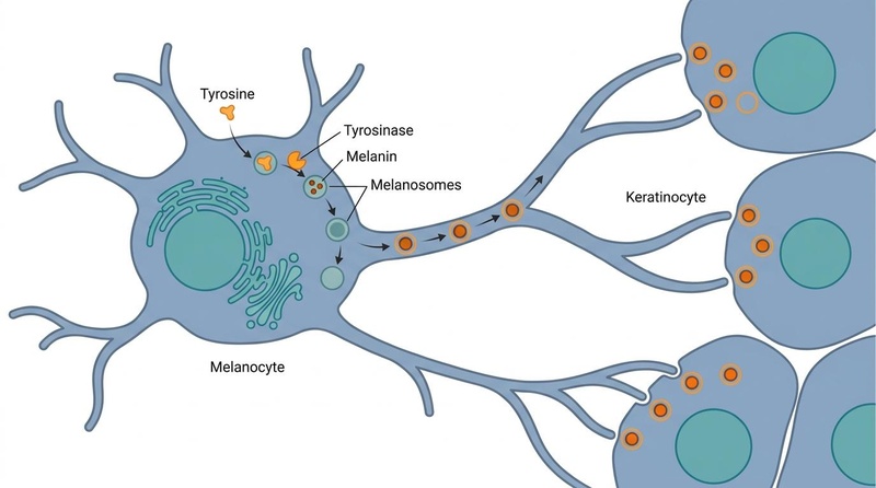
Hyperpigmentation (PIH, melasma, solar lentigines) is primarily driven by melanogenesis, the complex biochemical process of melanin synthesis within specialized cells called melanocytes. When skin experiences inflammation (from acne, injury, or irritation) or excessive UV exposure, melanocytes are stimulated to produce more melanin. This melanin is then packaged into organelles called melanosomes, which are subsequently transferred to neighboring keratinocytes (the primary cells of the epidermis). The accumulation of excess melanin in the epidermis and/or dermis manifests as visible dark spots on the skin.
::: .fold
¶ Image Metadata: Melanogenesis Pathway
- Visual Plan: Clean, elegant biomedical illustration detailing the process of melanogenesis within a melanocyte and the subsequent transport of melanin-containing melanosomes to surrounding keratinocytes. It highlights the biochemical pathway where tyrosine and tyrosinase interact to produce melanin inside developing melanosomes, which then travel along the melanocyte's dendritic processes.
- Model ID:
gemini-3-pro-image(Nano Banana Pro-backed generation) - Prompt: "Create a detailed biomedical illustration of Biochemical pathway of melanogenesis inside a melanocyte. Show the conversion of tyrosine to melanin catalyzed by tyrosinase, and the transport of melanosomes along dendritic processes to neighboring keratinocytes, using a clean biomedical editorial style. Intracellular organelles and migrating melanosome vesicles. Balanced composition with soft anatomical/molecular abstraction. White/off-white background, slate-gray structure lines, muted blue and teal biological forms, subtle warm orange accents for emphasis. Minimal, elegant, encyclopedia-ready.. Use a BioRender-like style (avoid any "BioRender" text or logo) but make sure to incorporate a bright orange colour, possibly for details and highlights and keep the background white. Make it look as if it was to appear in Nature or Science. Do NOT include any text saying this is a Nature/Science image."
- Caption: "Figure: Melanogenesis Pathway. Tyrosinase catalyzes the conversion of tyrosine to melanin inside melanosomes, which are then transported along dendritic processes to adjacent keratinocytes."
- Alt Text: "Clean, elegant biomedical illustration of the melanogenesis pathway within a melanocyte, showing tyrosine conversion to melanin and melanosome transport along dendrites to keratinocytes."
- Placement: Under section
What Causes It->Pigment Formation (Melanogenesis) - QA State:
passed(Verified for scientific accuracy and stylistic alignment)
:::
- UV Radiation: A primary trigger for melanogenesis, leading to increased melanin production and darkening of existing pigment marks and exacerbating melasma [2:1].
- Inflammation: Cytokines and inflammatory mediators released during acne lesions or skin injury stimulate melanocytes, resulting in PIH [1:2].
- Hormonal Factors: Estrogen and progesterone can stimulate melanocytes, contributing to melasma, particularly during pregnancy or with oral contraceptive use [2:2].
¶ Collagen Destruction and Dysregulation from Acne Inflammation
Acne scars, particularly atrophic scars, result from abnormal wound healing processes following deep inflammatory acne lesions.
- Inflammation: Severe or prolonged acne inflammation can damage collagen and elastic fibers in the dermis.
- Enzymatic Degradation: Inflammatory cells release enzymes (matrix metalloproteinases) that break down collagen more rapidly than it can be replaced.
- Impaired Collagen Synthesis: During the healing process, there can be inadequate synthesis of new collagen, or the new collagen fibers may be disorganized, leading to depressed areas (atrophic scars) [3:6].
- Excessive Collagen Deposition: Conversely, in hypertrophic and keloid scars, there is an overproduction and accumulation of collagen, leading to raised, thickened scar tissue [3:7].
¶ Starter Protocol (Topical Agents)
For initial management and maintenance, a consistent regimen of topical agents is crucial.
- Broad-Spectrum Sunscreen (SPF 50+): Absolutely non-negotiable. Apply daily, year-round, even indoors or on cloudy days. Sunscreen prevents new hyperpigmentation, deepens existing pigment, and protects against UV-induced collagen breakdown. Look for formulations with physical blockers (zinc oxide, titanium dioxide) and iron oxides for visible light protection, especially for melasma [2:3].
- Topical Retinoids (e.g., Adapalene, Tretinoin): Increase cell turnover, promoting the exfoliation of pigmented cells and preventing clogged pores. They also stimulate collagen production, which can modestly improve superficial atrophic scars. Start with lower strengths a few times a week and gradually increase frequency as tolerated to minimize irritation.
- Vitamin C (L-Ascorbic Acid): A potent antioxidant that inhibits tyrosinase activity (an enzyme critical for melanin production) and brightens the skin. It also supports collagen synthesis. Use a stable formulation in the morning.
- Niacinamide (Vitamin B3): Reduces the transfer of melanosomes from melanocytes to keratinocytes, helping to prevent pigment from reaching the skin's surface. It also has anti-inflammatory properties and can improve skin barrier function.
- Azelaic Acid (10-20%): A natural dicarboxylic acid with anti-inflammatory, antibacterial, and tyrosinase-inhibiting properties. Highly effective for both PIH and melasma, and generally well-tolerated, even in sensitive skin or skin of color [1:3][16].
- Salicylic Acid (BHA): A beta-hydroxy acid that exfoliates inside pores, making it effective for active acne and helping to reduce superficial PIH by promoting cell turnover.
¶ Stronger Add-On Path
When the starter protocol is insufficient, consider incorporating more potent agents under clinical guidance.
- Hydroquinone (2-4%): A powerful tyrosinase inhibitor considered the gold standard for reducing hyperpigmentation. Due to potential side effects like irritation, contact dermatitis, and rare exogenous ochronosis with prolonged use, it is typically used for short durations (3-6 months) and often cycled with non-hydroquinone agents [17].
- Cysteamine Cream (5%): A non-hydroquinone topical depigmenting agent with comparable efficacy to hydroquinone for melasma and PIH, and a favorable safety profile for all skin types. It works by reducing melanin production. Apply for short contact (15-20 minutes) before washing off [5:1][4:1].
- Tranexamic Acid (Oral or Topical):
- Oral (typically 250-500mg daily): Highly effective for melasma, particularly refractory cases, by inhibiting plasminogen activator, which reduces melanin synthesis and vascularity [7:1][8:1]. Requires medical supervision due to potential contraindications (e.g., history of thromboembolic events).
- Topical: Can be used as an adjunct to reduce hyperpigmentation and erythema.
- Higher Strength Retinoids (e.g., Tretinoin): Prescription-strength retinoids provide more potent cell turnover and collagen stimulation, offering enhanced benefits for PIH, fine lines, and superficial scarring. Use under medical guidance due to increased potential for irritation.
- Home Microneedling (Dermarolling/Dermastamping): Can enhance the penetration of topical agents and stimulate some collagen remodeling for very superficial scars. Use with caution to avoid infection or exacerbating PIH, especially in darker skin types. Strict sterilization and appropriate needle depth (e.g., 0.25mm-0.5mm) are essential.
¶ Clinical/Procedure Path
For more significant or recalcitrant blemishes, pigment marks, and acne scars, in-office procedures performed by a qualified clinician offer advanced solutions.
- Chemical Peels:
- Superficial Peels (e.g., Glycolic Acid, Lactic Acid, Salicylic Acid): Promote exfoliation of the outermost skin layers, effective for mild PIH, superficial acne scars, and improving overall skin texture. Generally safe for all skin types with proper formulation and technique.
- Medium-Depth Peels (e.g., TCA 20-35%): Penetrate deeper to address more significant pigmentation and textural irregularities. Require greater caution, especially in skin of color, due to increased risk of PIH. Meticulous pre- and post-peel care is essential [18].
- Microneedling (Collagen Induction Therapy): Utilizes a device with fine needles to create controlled micro-injuries in the skin, stimulating collagen and elastin production. Effective for rolling and shallow boxcar scars, and can improve PIH. Multiple sessions are needed.
- Radiofrequency (RF) Microneedling: Combines microneedling with radiofrequency energy, delivering heat into the deeper dermis to enhance collagen tightening and remodeling, making it more effective for rolling scars and overall texture improvement.
- Subcision: A minor surgical procedure particularly effective for rolling acne scars. A needle or cannula is inserted under the scar to physically break the fibrous bands that tether the skin down, releasing the depression [11:1][12:1]. Often combined with dermal fillers or platelet-rich plasma (PRP) for sustained volume and healing [12:2].
- Laser Resurfacing (e.g., Fractional CO2, Erbium:YAG):
- Ablative Lasers: Create microscopic columns of thermal injury, vaporizing tissue and stimulating significant collagen remodeling. Highly effective for deep atrophic scars but require significant downtime and carry a higher risk of PIH, especially in darker skin tones [19][20].
- Non-Ablative Fractional Lasers: Target water in the skin to create controlled thermal injury without ablating the surface, leading to less downtime but requiring more sessions. Improve texture and pigmentation.
- IPL (Intense Pulsed Light) / Q-switched Lasers / Picosecond Lasers: Energy-based devices that target melanin (for PIH, melasma, solar lentigines) or hemoglobin (for PIE). Careful selection of parameters and patient skin type is paramount to avoid adverse effects like PIH exacerbation [19:1]. Pulsed Dye Lasers (PDL) are gold standard for PIE [13:1].
- TCA CROSS (Chemical Reconstruction Of Skin Scars): Focal application of high-concentration (70-100%) trichloroacetic acid directly to the base of ice pick and deep boxcar scars. This creates a localized chemical burn that stimulates significant collagen remodeling and scar elevation [10:1][3:8]. Requires precise technique and multiple sessions.
¶ What Usually Fails
Understanding ineffective or harmful approaches is as important as knowing what works.
- DIY Lemon Juice or Other "Natural" Bleaching Agents: Often ineffective and can cause significant skin irritation, phototoxicity, and even worsen hyperpigmentation due to their acidic nature and lack of standardized concentration.
- Aggressive Scrubbing or Physical Exfoliants: Can compromise the skin barrier, leading to increased inflammation, which in turn can exacerbate PIE and PIH, and potentially worsen acne.
- Weak Over-The-Counter (OTC) Creams for Deep Scars: While some OTC products can help with superficial texture or pigment, they are generally insufficient to address true atrophic or hypertrophic acne scars, which require medical procedures.
- Ignoring Active Acne: Attempting to treat scars or pigment marks while active acne is still present is counterproductive, as new lesions will continue to create new marks.
- Inconsistent Sun Protection: The most common reason for treatment failure in pigmentary disorders. Without strict daily sun protection, hyperpigmentation will rebound and worsen.
- Expecting Overnight Results: Treatments for scars and pigment marks require patience and consistency over several weeks to months to see noticeable improvement.
¶ Tracking Plan
Objective tracking of progress helps evaluate treatment efficacy and guide adjustments.
- Standardized Clinical Photography: Regular (e.g., every 4-8 weeks) high-quality photographs taken under consistent lighting, angle, and distance are invaluable for documenting visual changes over time.
- Wood's Lamp Evaluation: A diagnostic tool that uses ultraviolet light to visualize pigment changes. Epidermal pigmentation appears enhanced under Wood's lamp, while dermal pigmentation shows little change. This can help differentiate superficial from deep pigmentary lesions and track lightening.
- Scar Texture Logs/Assessment Scales: Clinicians may use objective scales like the Global Acne Scarring Classification or subjective patient-reported outcome measures (e.g., visual analogue scales, quality of life questionnaires) to track changes in scar depth, texture, and overall appearance. Patients can maintain personal "scar diaries" to note perceived changes.
- MASI Score (Melasma Area and Severity Index): A validated clinical tool used by dermatologists to quantify the severity of melasma. While complex for self-assessment, simplified versions or regular clinical evaluation can track melasma improvement [2:4].
- Topical Treatment Adherence Logs: Keeping a record of product application (what, when, how often) helps ensure consistency and identify potential gaps in adherence.
¶ Safety and Red Flags
Awareness of potential risks and knowing when to seek professional medical advice are critical for safe and effective management.
¶ General Safety Considerations
- Strict Sun Protection (SPF 50+): Paramount during and after any treatment for pigmentary issues or scarring. UV exposure is the leading cause of PIH and melasma exacerbation [2:5].
- Skin of Color (Fitzpatrick Types IV-VI): Individuals with darker skin are at a significantly higher risk of developing post-inflammatory hyperpigmentation (PIH) or, less commonly, hypopigmentation (loss of pigment) following aggressive procedures like certain laser treatments, deeper chemical peels, or even microneedling if parameters are not meticulously chosen [19:2][20:1][18:1]. It is crucial to use lower energy settings, appropriate wavelengths (e.g., Nd:YAG lasers for darker skin), and follow strict pre- and post-treatment protocols.
- Hydroquinone Use: Continuous application of hydroquinone should be limited to 3-6 months to minimize the rare but serious risk of exogenous ochronosis (irreversible bluish-black discoloration of the skin) [17:1]. Cycle hydroquinone with non-hydroquinone lightening agents.
- Oral Tranexamic Acid: Contraindicated in patients with a personal or family history of thromboembolic disease (e.g., deep vein thrombosis, pulmonary embolism), or other risk factors for blood clots. A thorough medical history and clinician consultation are mandatory before use.
- Pregnancy and Lactation: Many active topical ingredients (e.g., retinoids, hydroquinone) and most in-office procedures are contraindicated or require careful consideration during pregnancy and breastfeeding.
- Active Skin Conditions: Avoid most aesthetic procedures on areas with active infections (bacterial, viral, fungal), open wounds, or other inflammatory skin conditions (e.g., severe rosacea flare) to prevent worsening the condition or introducing complications.
¶ Red Flags (When to Talk to a Clinician)
- Lack of Response or Worsening: If blemishes, pigment marks, or scars are not responding to consistent home care, or if they are worsening despite adherence to treatment and rigorous sun protection.
- New or Rapidly Changing Lesions: Any new skin lesion that is unusually shaped, rapidly growing, bleeding, itching intensely, or painful should be promptly evaluated by a dermatologist to rule out malignancy or other serious conditions.
- Signs of Infection: Increased redness, swelling, warmth, pain, pus, or fever in treated areas after a procedure or topical application.
- Severe Allergic Reactions: Widespread rash, hives, severe itching, swelling of the face or throat, or difficulty breathing after using a new product.
- Unusual Scar Progression: If hypertrophic scars or keloids are rapidly enlarging, becoming increasingly painful, or interfering with normal function (e.g., joint movement).
- Significant Psychological Distress: If skin concerns are leading to substantial anxiety, depression, social withdrawal, or a significant negative impact on quality of life, professional psychological support may be beneficial alongside dermatological treatment.
¶ FAQs
¶ Can acne scars go away naturally?
Most true acne scars, which are permanent textural changes (atrophic or hypertrophic), do not resolve naturally without intervention. Post-inflammatory erythema (red marks) and post-inflammatory hyperpigmentation (dark marks) can fade over months to years, but treatment can significantly accelerate and improve this process.
¶ Is microneedling good for all acne scars?
Microneedling is most effective for improving rolling and shallow boxcar atrophic scars by stimulating collagen production. It is less effective for deep ice pick scars or raised hypertrophic/keloid scars, which often require more targeted treatments such as TCA CROSS, subcision, or intralesional injections.
¶ How long does it take for PIH to fade?
Post-inflammatory hyperpigmentation can take several months to years to fade naturally, depending on its depth, the individual's skin type, and sun exposure. With consistent use of depigmenting agents and strict sun protection, significant improvement can often be observed within 8-12 weeks.
¶ Can chemical peels be used on darker skin tones?
Yes, certain chemical peels can be safely and effectively used on darker skin tones (Fitzpatrick IV-VI). However, careful selection of the acid type, concentration, and peel depth is crucial. Superficial peels (e.g., glycolic, lactic, salicylic acids) are generally safer, while medium-depth peels carry a higher risk of post-inflammatory hyperpigmentation if not performed by an experienced professional with appropriate pre- and post-peel care [18:2].
¶ What is the difference between PIE and PIH?
PIE (Post-Inflammatory Erythema) refers to persistent red or purple marks left after inflammatory acne, caused by damaged or dilated blood vessels. PIH (Post-Inflammatory Hyperpigmentation) refers to brown, black, or gray spots resulting from excess melanin production after inflammation. PIE is primarily treated with vascular-targeting therapies like pulsed dye lasers, while PIH responds to melanin-inhibiting and exfoliating agents.
¶ References
Zaher H, El-Komy M, Hassan S, Abozeid T. Effects of 15% Azelaic Acid Gel in the Management of Post-Inflammatory Erythema and Post-Inflammatory Hyperpigmentation in Acne Vulgaris. Clin Cosmet Investig Dermatol. 2024 May 13;17:663-671. https://pmc.ncbi.nlm.nih.gov/articles/PMC11116308/ ↩︎ ↩︎ ↩︎ ↩︎
Rerksirattana P, Pothacharoen P, Tuchinda P, Imhof M, Leeyaphan C, Tanzi EL, Wulkan A, Bacci PA, Gold MH, Goodman G, Landau M, Naito Z, Vasanop V, Vachiramon V, Viyoch J. Delphi consensus on melasma management by international experts and pigmentary disorders society. J Eur Acad Dermatol Venereol. 2021 Oct;35(10):2064-2078. https://pubmed.ncbi.nlm.nih.gov/40996222/ ↩︎ ↩︎ ↩︎ ↩︎ ↩︎ ↩︎
Zhang M, Liu C, Zhang S, Chu R, Ren X. Advances in the treatment of acne scars. Front Med (Lausanne). 2025 Aug 29;12:1643035. https://pmc.ncbi.nlm.nih.gov/articles/PMC12425707/ ↩︎ ↩︎ ↩︎ ↩︎ ↩︎ ↩︎ ↩︎ ↩︎ ↩︎
Mansouri P, Farshi S, Hashemi Z, Kasraee B. Evaluation of the efficacy of cysteamine 5% cream in the treatment of epidermal melasma: a randomized double-blind placebo-controlled trial. Br J Dermatol. 2015 Jul;173(1):209-17. https://academic.oup.com/bjd/article-abstract/173/1/209/6616816 ↩︎ ↩︎
Li M, Wang Y, Hu X, Wei Y, Wei Y. Efficacy and safety of cysteamine 5% cream for the management of melasma: a systematic review and meta-analysis of randomized controlled trials. J Cosmet Dermatol. 2024 Sep;23(9):2838-2850. https://pubmed.ncbi.nlm.nih.gov/39673630/ ↩︎ ↩︎
Al-Sunaidi N, Al-Mutairi N, Ali Y. Comparison of the efficacy of cysteamine 5% cream and hydroquinone 4%/ascorbic acid 3% combination cream in the treatment of epidermal melasma. J Cosmet Dermatol. 2022 Jun;21(6):2614-2620. https://pubmed.ncbi.nlm.nih.gov/35510765/ ↩︎
Liang B, Chen X, Liang F, Zhang X, Li Z. Tranexamic acid as a therapeutic option for melasma management: meta-analysis and systematic review of randomized controlled trials. J Cosmet Dermatol. 2024 Aug 29. https://pubmed.ncbi.nlm.nih.gov/38843906/ ↩︎ ↩︎
Na J, Kim SY, Kim SM, Ko JY, Kim H, Shin MK. Tranexamic Acid for Hyperpigmentation Disorders: A Literature Review on Efficacy and Safety in Melasma and PIH. J Clin Med. 2023 Dec 25;13(1):96. https://pmc.ncbi.nlm.nih.gov/articles/PMC12848551/ ↩︎ ↩︎
Kircik LH. Efficacy and safety of azelaic acid (AzA) gel 15% in the treatment of post-inflammatory hyperpigmentation and acne: a 16-week, baseline-controlled study. J Drugs Dermatol. 2011 May;10(5):586-90. https://pubmed.ncbi.nlm.nih.gov/21637899/ ↩︎
Bhardwaj D, Khunger N. An Assessment of the Efficacy and Safety of CROSS Technique with 100% TCA in the Management of Ice Pick Acne Scars. J Cutan Aesthet Surg. 2010 Jul;3(2):93-6. https://pmc.ncbi.nlm.nih.gov/articles/PMC2956965/ ↩︎ ↩︎
Al-Dhalimi MA, Arnoos AA. Subcision for treatment of rolling acne scars in Iraqi patients: a clinical study. J Cosmet Laser Ther. 2012 Jun;14(3):149-53. https://pubmed.ncbi.nlm.nih.gov/22672279/ ↩︎ ↩︎
Ahramiyanpour N, Khosravi-Shahi M. Subcision in acne scarring: A review of clinical trials. J Cosmet Dermatol. 2023 Dec;22(12):3211-3220. https://pubmed.ncbi.nlm.nih.gov/36315903/ ↩︎ ↩︎ ↩︎
Kalantari Y, Aghazadeh S, Khosravi H, Kalantari N. Post-acne erythema treatment: A systematic review of the literature. J Cosmet Dermatol. 2022 Feb;21(2):494-501. https://pubmed.ncbi.nlm.nih.gov/35076997/ ↩︎ ↩︎
Bae-Harboe YS, Graber EM. Easy as PIE (Postinflammatory Erythema). J Clin Aesthet Dermatol. 2013 Sep;6(9):36-7. https://pmc.ncbi.nlm.nih.gov/articles/PMC3780804/ ↩︎
Zhu X, Song X, Zeng Z, Li S, Fu X, Zeng M, Li Z. Optimal treatment options for acne scars in patients with historic acne: a systematic review and network meta-analysis. BMC Med. 2024 Jul 12;22(1):285. https://pmc.ncbi.nlm.nih.gov/articles/PMC12515001/ ↩︎
Alnajjar Z, Alshami A, Aldallal A, Alkhunaizi S, Alamri M, Almutairi S, Alamri F. Azelaic Acid Versus Hydroquinone for Managing Patients With Melasma: Systematic Review and Meta-Analysis of Randomized Controlled Trials. Cureus. 2023 Jul 12;15(7):e41743. https://pmc.ncbi.nlm.nih.gov/articles/PMC10339666/ ↩︎
Gupta AK, Waddell LS, Perera P, Perera N. Melasma Treatment: An Evidence-Based Review. Am J Clin Dermatol. 2020 Oct;21(5):669-682. https://pubmed.ncbi.nlm.nih.gov/31802394/ ↩︎ ↩︎
Garelick E, Pohani P, Pousti S, Chen SC, Lee PK. Chemical Peels in Skin of Color: A Scoping Review of Safety, Efficacy. Lehigh Valley Health Network. 2023. https://scholarlyworks.lvhn.org/education-medical-education/183/ ↩︎ ↩︎ ↩︎
Alexis AF, Blackcloud P, Bivens MM, Burnett MA, Callender VD, Chwalek J, Fewkes SM, Fitzpatrick R, Friedmann DP, Gold MH, Grimes PE, Kroumpouzos G, Lambros V, Layton AM, Lobl M, Long D, Luhn S, Mayhew C, McGill-Rodgers P, Mehta V, Monheit GD, Moyer J, Patel U, Photiades P, Sadick N, Shaub A, Taub AF, Werschler WP. Noninvasive Cosmetic Treatments for Fitzpatrick IV–VI: A Narrative Review of Safety and Efficacy Guidelines. J Clin Aesthet Dermatol. 2023 Dec;16(12):12-24. https://pmc.ncbi.nlm.nih.gov/articles/PMC13012588/ ↩︎ ↩︎ ↩︎
Ho D, Trinh N, Sivamani RK, et al. Laser Fitzpatrick Skin Type Recommendations. [Updated 2024 Jun 29]. In: StatPearls [Internet]. Treasure Island (FL): StatPearls Publishing; 2024 Jan-. https://www.ncbi.nlm.nih.gov/books/NBK557626/ ↩︎ ↩︎