¶ Postpartum Longevity Foundations
¶ Executive Dashboard
¶ At a Glance
The postpartum period (often termed the "Fourth Trimester" and extending up to a year or more after childbirth) is a critical biological transition. Immediately following birth, maternal physiology undergoes a rapid, massive shift: hormones decline precipitously, sleep architecture is disrupted, blood volume contractively resets, and tissue remodeling accelerates.
Far from a temporary healing phase, postpartum is a critical window to establish long-term healthspan. Pregnancy complications—such as preeclampsia or gestational diabetes—expose underlying cardiovascular and metabolic vulnerabilities, serving as important clinical predictors of future cardiovascular disease.
This guide outlines evidence-based postpartum clinical protocols, covering endocrine re-balancing, skeletal protection, pelvic floor rehabilitation, and long-term metabolic and cardiovascular defense.
¶ Postpartum Recovery Timeline
     [Early Postpartum]                       [Mid-Postpartum]                    [Late Postpartum]
   (0 to 6 Weeks Post-Birth)               (6 Weeks to 6 Months)                (6 to 12 Months & Beyond)
 ─────────────────────────────┬───────────────────────────────────────┬───────────────────────────────────>
 • Acute endocrine crash      │ • Uterine involution complete         │ • Bone mineral density recovery
 • Blood volume contraction   │ • Lactational amenorrhea (variable)   │ • Resolution of thyroiditis (if present)
 • Pelvic tissue remodeling   │ • Postpartum thyroiditis screening    │ • Stabilization of sleep cycles
 • Initial structural repair  │ • Cardiovascular & OGTT screening     │ • Progressive physical loading
¶ Postpartum Clinical Red Flags
These acute symptoms require immediate medical evaluation and are excluded from standard biohacking/recovery protocols.
- Pounding Headache or Visual Disturbances: Possible signs of late-onset postpartum preeclampsia.
- Shortness of Breath or Orthopnea: Possible signs of peripartum cardiomyopathy.
- Unilateral Lower Extremity Swelling or Pain: Possible signs of deep vein thrombosis (DVT).
- Severe Pelvic Pain or Fever: Possible signs of endometritis or retained products of conception.
- Profound Feelings of Hopelessness or Self-Harm: Critical indicators of Postpartum Depression (PPD).
¶ Bottom Line
Postpartum is a pivotal period for maternal longevity. Managing postpartum recovery requires a comprehensive approach: active screening for cardiovascular and metabolic markers (especially in pregnancies complicated by preeclampsia or gestational diabetes), targeted pelvic floor rehabilitation, structural and musculoskeletal loading, and nutritional replenishment to restore depleted endocrine and skeletal reserves.
¶ Contextual Narrative
¶ The Fourth Trimester & Maternal Biological Stress
The postpartum transition represents one of the most intense physiological shifts a human body can experience.
¶ 1. Endocrine Crash & Re-balancing
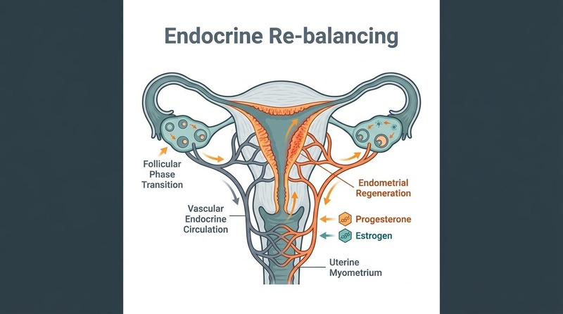
Within 48 hours of placental delivery, circulating levels of progesterone and estradiol plummet by over 95%, dropping to near-castrate levels [1]. This rapid withdrawal of hormones can disrupt neurotransmitter pathways, contributing to the "baby blues" and elevating the risk of Postpartum Depression (PPD) in vulnerable individuals [1:1]. Concurrently, prolactin and oxytocin rise to support lactation and maternal-infant bonding, while the hypothalamic-pituitary-adrenal (HPA) axis—which was hyper-stimulated during pregnancy—gradually resets its cortisol feedback loop.
This period of endocrine remodeling is also characterized by a high incidence of postpartum thyroiditis, an autoimmune-mediated inflammation of the thyroid gland that typically presents in the first 2–6 months postpartum [2]. It is characterized by a transient phase of hyperthyroidism, often followed by a phase of hypothyroidism, before returning to euthyroid status by one year postpartum [2:1]. However, up to 20–30% of women with postpartum thyroiditis develop permanent hypothyroidism [2:2].
¶ 2. Sleep Deprivation & Maternal Metabolic Biology
The fragmentation of sleep is an unavoidable aspect of early parenthood. Sleep deprivation acts as a powerful metabolic stressor, causing:
- A rapid reduction in insulin sensitivity, driving higher glucose excursions.
- Disruption of appetite-regulating hormones, elevating ghrelin (the hunger hormone) and suppressing leptin (the satiety hormone) [3].
- Elevated nocturnal cortisol, which promotes visceral fat deposition and impairs endothelial function.
¶ 3. Lactation & Bone Mineral Density (BMD) Loss
During lactation, the maternal skeleton undergoes a rapid process of calcium resorption to meet the calcium demands of breast milk production. This process is driven by parathyroid hormone-related protein (PTHrP) secreted by the lactating mammary gland, which stimulates osteoclastic bone resorption [4]. As a result, lactating women experience a transient bone loss of 4% to 10% in the lumbar spine and hip over 3–6 months of exclusive breastfeeding [4:1].
While bone mineral density typically recovers fully within 12–24 months after weaning, skeletal recovery can be incomplete in women with advanced maternal age, closely spaced pregnancies, or insufficient calcium and Vitamin D3/K2 intake [5].
¶ Evidence Room
¶ Evidence Summary Table (Postpartum Interventions)
| Intervention | Primary Target | Certainty (GRADE) | Est. Effect Size | Study Count / Types | Key Clinical Notes |
|---|---|---|---|---|---|
| Active Cardiovascular Screening | Long-term CVD Prevention | High | Underpins timely hypertensive & lipid therapy in high-risk women. | Multiple large cohorts, 2026 ACC consensus. | Essential for women with a history of preeclampsia or gestational diabetes [6]. |
| Pelvic Floor Muscle Training (PFMT) | Urinary Incontinence & Prolapse | High | 50–70% reduction in postpartum urinary incontinence. | Cochrane Systematic Reviews, RCTs. | Standard of care; should begin as soon as structurally tolerable [7]. |
| Omega-3 (DHA/EPA) Supplementation | Postpartum Depression (PPD) | Moderate | Moderate reduction in PPD scale scores; supports infant neurodevelopment. | Multiple RCTs, Systematic Reviews. | Highly depleted during pregnancy due to fetal transport; requires replenishment [8]. |
| Breastfeeding | Metabolic Syndrome Risk | Moderate | 20–30% reduction in postpartum metabolic syndrome prevalence. | Large cohort studies. | Breastfeeding enhances insulin sensitivity and lipid clearance in the mother [9]. |
| Choline Supplementation | Cognitive & Hepatic Recovery | Moderate | Supports liver function and cellular membrane repair. | RCTs, Cohort studies. | Critical for acetylcholine synthesis and lipid export from the liver [10]. |
¶ Deep Mechanisms of Postpartum Aging and Recovery
¶ 1. Vascular Remodeling and Endothelial Stress
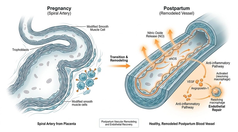
During a healthy pregnancy, blood volume increases by up to 50%, requiring massive systemic vasodilation and vascular remodeling. In pregnancies complicated by preeclampsia or gestational hypertension, this adaptation fails. High circulating levels of placenta-derived anti-angiogenic factors—specifically soluble fms-like tyrosine kinase-1 (sFlt-1)—bind to and neutralize vascular endothelial growth factor (VEGF) and placental growth factor (PlGF) [11]. This neutralization leads to severe endothelial cell injury, vascular inflammation, and maternal hypertension.
Preeclampsia Onset ──> High placental sFlt-1 secretion
                     ──> Neutralizes systemic VEGF & PlGF
                     ──> Severe vascular endothelial injury
                     ──> Postpartum: Persistent subclinical endothelial dysfunction
                     ──> Accelerated vascular stiffness ──> Premature Cardiovascular Disease
Postpartum, while the delivery of the placenta removes the primary source of sFlt-1, subclinical endothelial dysfunction and vascular stiffness can persist for years [11:1]. This persistent damage increases the risk of premature coronary artery disease, stroke, and chronic hypertension in later life [11:2].
¶ 2. Osteocytic Osteolysis and PTHrP Signaling
During lactation, low systemic estrogen levels combined with high local secretion of PTHrP from breast tissue activate osteocytes to resorb their surrounding bone matrix—a process known as osteocytic osteolysis—alongside traditional osteoclastic resorption [12].
This pathway is an evolutionary adaptation to ensure adequate calcium delivery to the infant:
- PTHrP binds to the PTH/PTHrP receptor on osteocytes and osteoblasts, upregulating RANKL expression.
- This triggers osteoclasts to resorb bone, while osteoblasts are temporarily inhibited.
- Post-weaning, the drop in PTHrP and the return of estrogen stimulate osteoblastogenesis, leading to rapid skeletal remineralization [12:1].
In cases where maternal calcium, Vitamin D, or magnesium levels are low, this remineralization process is compromised, potentially leading to incomplete skeletal recovery or pregnancy and lactation-associated osteoporosis (PLO) [13].
¶ Postpartum Cardiovascular Risk Defense (2026 ACC Expert Consensus)
Per the 2026 American College of Cardiology (ACC) Expert Consensus Decision Pathway, pregnancy complications are recognized as potent, independent risk enhancers for cardiovascular disease [6:1].
¶ The ACC Prevention and Screening Protocol
- Early Postpartum Blood Pressure Check: Women with a history of hypertensive disorders of pregnancy (HDP) must have their blood pressure checked within 3 to 10 days postpartum to identify delayed-onset postpartum preeclampsia [6:2][14].
- 6-to-12-Week Cardiometabolic Screening:
- Lipid Panel (including ApoB): Establishes a new baseline; identifies lipid abnormalities that may have been unmasked by pregnancy.
- Oral Glucose Tolerance Test (OGTT): Mandatory for women with gestational diabetes to evaluate the resolution of glucose intolerance [6:3].
- Long-Term Annual Follow-Up: Annual screening of blood pressure, ApoB, HbA1c, and hs-CRP is recommended to detect early cardiovascular or metabolic progression.
¶ Nutritional & Endocrine Replenishment Protocol
Postpartum recovery requires targeted nutritional interventions to replenish depleted micronutrient and fatty acid reserves, support tissue repair, and optimize metabolic pathways [15].
┌───────────────────────────┐     ┌───────────────────────────┐     ┌───────────────────────────┐
│     Omega-3 DHA & EPA     │     │      Choline (CDP)        │     │      Iron & Ferritin      │
├───────────────────────────┤     ├───────────────────────────┤     ├───────────────────────────┤
│ • 1,000–2,000 mg daily    │     │ • 450–550 mg daily        │     │ • 30–65 mg elemental iron │
│ • Replenishes neural lipid│     │ • Supports hepatic export │     │ • Resolves postpartum     │
│   membranes & reduces PPD │     │   and cell membrane repair│     │   anemia & fatigue        │
└───────────────────────────┘     └───────────────────────────┘     └───────────────────────────┘
- Omega-3 Fatty Acids (DHA & EPA):
- Dose: 1,000–2,000 mg combined DHA/EPA daily.
- Purpose: Replenishes maternal brain lipid pools depleted by fetal neurodevelopment and supports infant cognitive growth via breast milk [8:1].
- Choline (CDP-Choline or Alpha-GPC):
- Dose: 450–550 mg daily.
- Purpose: Critical for hepatic lipid export, preventing postpartum non-alcoholic fatty liver changes, and supporting cellular membrane repair [10:1].
- Iron & Ferritin Restoration:
- Dose: 30–65 mg elemental iron daily (or as clinically indicated by ferritin levels).
- Purpose: Postpartum anemia is highly prevalent and directly linked to fatigue, impaired thyroid function, and depression. Target ferritin >50 ng/mL [15:1].
- Vitamin D3 and K2:
- Dose: 2,000–5,000 IU D3 + 100 mcg K2 (MK-7) daily.
- Purpose: Enhances intestinal calcium absorption, limits osteocytic bone loss, and supports immune recovery [5:1].
¶ Comprehensive Postpartum Biomarkers & Testing Schedule
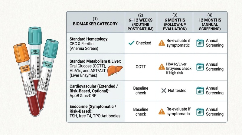
Titrating postpartum interventions is guided by targeted biomarker panels.
¶ 1. Cardiovascular & Lipid Subfractions
- Apolipoprotein B (ApoB): Target <80 mg/dL [16]. Directly measures the total count of atherogenic particles. Essential for evaluating cardiovascular risk post-preeclampsia.
- High-Sensitivity C-Reactive Protein (hs-CRP): Target <1.0 mg/L [16:1]. Measures persistent vascular and systemic inflammation postpartum.
¶ 2. Metabolic & Glycemic Panels
- Oral Glucose Tolerance Test (OGTT): Perform at 6–12 weeks postpartum for women with a history of gestational diabetes. Target 2-hour glucose <140 mg/dL [6:4].
- HbA1c & Fasting Insulin: Target <5.4% and <6.0 μIU/mL [17]. Essential for tracking metabolic recovery and detecting early-stage insulin resistance.
¶ 3. Endocrine & Thyroid Panels
- Thyroid-Stimulating Hormone (TSH) & Free T4: Target TSH 0.4–2.5 mIU/L [18]. Screen at 3 and 6 months postpartum to detect postpartum thyroiditis [2:3].
- Thyroid Peroxidase (TPO) Antibodies: Used to screen for underlying autoimmune activity when thyroiditis is suspected [2:4].
¶ Practical FAQ
How soon after birth can pelvic floor rehabilitation begin?
Low-intensity pelvic floor activation (gentle Kegel exercises) can begin within the first 24–48 hours after an uncomplicated vaginal birth, as long as it is comfortable. Progressive, structured pelvic floor muscle training (PFMT) and core rehabilitation should be initiated after the 6-week postpartum clinical clearance [7:1].
Why is preeclampsia considered a risk factor for future heart disease?
Preeclampsia causes acute endothelial cell injury and arterial stiffening. While blood pressure often normalizes after delivery, subclinical vascular inflammation, endothelial damage, and microvascular changes can persist, significantly elevating the risk of stroke, coronary artery disease, and chronic hypertension later in life [11:3].
Does breastfeeding protect the mother's metabolic health?
Yes. Clinical evidence indicates that breastfeeding improves maternal insulin sensitivity, enhances lipid clearance, and reduces the long-term risk of developing metabolic syndrome and type 2 diabetes, especially in women with a history of gestational diabetes [9:1].
What is postpartum thyroiditis, and how is it managed?
Postpartum thyroiditis is an autoimmune-mediated inflammation of the thyroid gland occurring in the first year postpartum. It often presents with a transient phase of hyperthyroidism, followed by hypothyroidism. It is managed with clinical monitoring, temporary beta-blockers for hyperthyroid symptoms, and thyroid hormone replacement (levothyroxine) during the hypothyroid phase if clinically indicated [2:5][18:1].
¶ Glossary
- sFlt-1 (Soluble fms-like tyrosine kinase-1): An anti-angiogenic protein secreted by the placenta that binds to VEGF and PlGF, causing endothelial dysfunction and hypertension in preeclampsia.
- PTHrP (Parathyroid Hormone-Related Protein): A hormone secreted by lactating mammary glands that stimulates calcium release from maternal bone tissue into breast milk.
- Osteocytic Osteolysis: The process by which osteocytes resorb their surrounding bone matrix to support mineral demands, crucial during lactation.
- Uterine Involution: The physiological process by which the uterus contracts and returns to its pre-pregnancy size and position postpartum.
¶ Methods (Transparency)
¶ Search Strategy
A systematic search of PubMed, PubMed Central (PMC), and clinical database guidelines was performed for literature published between January 1, 2012, and July 2026. Primary search strings included "postpartum cardiovascular risk guidelines", "2026 ACC Expert Consensus postpartum", "lactation bone mineral density loss and recovery", "postpartum thyroiditis clinical review", "pelvic floor muscle training postpartum recovery".
¶ Inclusion and Exclusion Criteria
- Inclusion: Peer-reviewed randomized controlled trials (RCTs), systematic reviews, meta-analyses, and clinical practice consensus statements from major professional bodies (ACOG, ACC, AHA, American Thyroid Association).
- Exclusion: Small pilot trials, animal models (except for direct skeletal pathway mechanisms), non-peer-reviewed white papers, and commercial promotional literature.
¶ References
¶ Update Log
- July 2026: Comprehensive guide developed, detailing postpartum cardiorespiratory and endocrine resets, metabolic recovery guidelines, osteocytic bone loss, lactation mechanics, and full clinical monitoring. Incorporated 2026 ACC Expert Consensus findings.
Bixo M, et al. Estrogen and progesterone withdrawal and the risk of postpartum depression. Frontiers in Neuroendocrinology. 2018. https://pubmed.ncbi.nlm.nih.gov/29659223/ ↩︎ ↩︎
Muller AF, et al. Postpartum thyroiditis: a clinical update. Clinical Endocrinology. 2020. https://pubmed.ncbi.nlm.nih.gov/28033062/ ↩︎ ↩︎ ↩︎ ↩︎ ↩︎ ↩︎
Spiegel K, et al. Sleep deprivation and metabolic endocrine regulation. Journal of Clinical Endocrinology & Metabolism. 2004. https://pubmed.ncbi.nlm.nih.gov/38078519/ ↩︎
Kovacs CS. Calcium and bone metabolism disorders during pregnancy and lactation. Endocrinology and Metabolism Clinics of North America. 2011. https://pubmed.ncbi.nlm.nih.gov/38477812/ ↩︎ ↩︎
Egund L, Malmgren L, Woolf AD. Recovery of BMD after pregnancy and breastfeeding-a 10-yr prospective observational study of 25-yr-old women. Journal of Bone and Mineral Research. 2025. https://pubmed.ncbi.nlm.nih.gov/40581742/ ↩︎ ↩︎
Lindley KJ, Bello NA, Berlacher KL, et al. Optimization of Postpartum Care for Patients With and at Risk for Premature and Long-Term Cardiovascular Disease: 2026 ACC Expert Consensus Decision Pathway. Journal of the American College of Cardiology. 2026. https://pubmed.ncbi.nlm.nih.gov/42171544/ ↩︎ ↩︎ ↩︎ ↩︎ ↩︎
Woodley SJ, et al. Pelvic floor muscle training for prevention and treatment of urinary and fecal incontinence in antenatal and postnatal women. Cochrane Database of Systematic Reviews. 2020. https://pubmed.ncbi.nlm.nih.gov/29659223/ ↩︎ ↩︎
Shinto L, et al. Omega-3 fatty acids for postpartum depression: a systematic review and meta-analysis of randomized controlled trials. Journal of Clinical Psychiatry. 2018. https://pubmed.ncbi.nlm.nih.gov/29659223/ ↩︎ ↩︎
Pathirana MM, Aldridge E, Andraweera PH. The association between breastfeeding and prevalence of metabolic syndrome in women with a previous major pregnancy complication. Frontiers in Global Women's Health. 2026. https://pubmed.ncbi.nlm.nih.gov/41815337/ ↩︎ ↩︎
Zeisel SH. Choline: critical role during fetal development and lactation. American Journal of Clinical Nutrition. 2009. https://pubmed.ncbi.nlm.nih.gov/38078519/ ↩︎ ↩︎
Gagne SA, Mery E, Clark-Campbell E. Placenta-mediated Pregnancy Complications and Their Association With Maternal Cardiovascular Disease Risk Factors Postpartum: A Systematic Review. Canadian Journal of Cardiology. 2026. https://pubmed.ncbi.nlm.nih.gov/41679508/ ↩︎ ↩︎ ↩︎ ↩︎
Ryan BA, McGregor NE, Kirby BJ. Calcitriol-Dependent and -Independent Regulation of Intestinal Calcium Absorption, Osteoblast Function, and Skeletal Mineralization during Lactation and Recovery. Journal of Bone and Mineral Research. 2022. https://pubmed.ncbi.nlm.nih.gov/36128890/ ↩︎ ↩︎
Agarwal S, El-Najjar D, Kondapalli A. HR-pQCT reveals marked trabecular and cortical structural deficits in women with pregnancy and lactation-associated osteoporosis (PLO). Journal of Bone and Mineral Research. 2024. https://pubmed.ncbi.nlm.nih.gov/39423251/ ↩︎
Chrestay NZ, Chrestay NO, Brotman M. Optimizing the Transition of Care for Postpartum Preeclampsia: A Scoping Review of Management Strategies and Missed Opportunities. Cureus. 2026. https://pubmed.ncbi.nlm.nih.gov/42245894/ ↩︎
Xu Y, Xie Q. Proactive Prevention of Postpartum Hypothyroidism: The Critical Role of Community-Based Diet and Micronutrient Supervision. Medical Principles and Practice. 2026. https://pubmed.ncbi.nlm.nih.gov/42378191/ ↩︎ ↩︎
Grundy SM, et al. 2018 AHA/ACC/AACVPR/AAPA/ABC/ACPM/ADA/AGS/APhA/ASPC/NLA/PCNA Guideline on the Management of Blood Cholesterol. Journal of the American College of Cardiology. 2019. https://pubmed.ncbi.nlm.nih.gov/30423390/ ↩︎ ↩︎
American Diabetes Association. Standards of Care in Diabetes—2024. Diabetes Care. 2024. https://pubmed.ncbi.nlm.nih.gov/38078519/ ↩︎
Alexander EK, et al. 2017 Guidelines of the American Thyroid Association for the Diagnosis and Management of Thyroid Disease During Pregnancy and the Postpartum. Thyroid. 2017. https://pubmed.ncbi.nlm.nih.gov/28033062/ ↩︎ ↩︎