¶ Breast Cancer Screening
| Primary Modality | 2D/3D Digital Mammography |
| Targeted Demographics | Asymptomatic individuals AFAB aged ≥40 |
| Screening Interval | Biennial or Annual (Risk-stratified) |
| Key Safety Concern | Overdiagnosis & False Positives |
| Downstream Cascade Risk | Surgical/core biopsies, patient anxiety |
| Access | Clinician referral or population screening |
Breast cancer is the second most common cancer and the second leading cause of cancer death among women in the United States [1]. Breast cancer screening aims to identify malignant changes in asymptomatic individuals before clinical signs emerge, advancing the time of diagnosis to a treatable stage. Within the context of a comprehensive Women's Longevity Guide and clinical management during the transition through Perimenopause and Menopause Longevity, breast cancer screening represents a cornerstone of proactive preventive medicine.
Landmark clinical trials and population registries demonstrate that routine mammography significantly reduces breast cancer-specific mortality [1:1][2][3]. However, screening is associated with distinct clinical trade-offs, including false-positive results, diagnostic cascades, psychological anxiety, and the identification of non-progressive cancers (overdiagnosis) [1:2][2:1].
¶ At a glance
- What people use it for: Early detection of malignant breast lesions in asymptomatic individuals to prevent disease progression and reduce breast cancer-specific mortality [1:3][2:2].
- Does it work? Yes. Large-scale randomized clinical trials and comparative simulation models demonstrate a reduction in breast cancer-specific mortality, ranging from approximately 15% in Cochrane and long-term Swedish trial overviews to 21% in standard Swedish trial overviews [4][5][3:1], and up to 25.4% to 41.7% in CISNET comparative modeling projections depending on screening intervals, starting ages, and age cohorts [6]. Furthermore, systematic reviews of clinical trials have not demonstrated a statistically significant reduction in all-cause mortality [4:1][3:2]. This is expected, as breast cancer-specific deaths represent only a small fraction of overall deaths in these cohorts, making trials under-designed to detect an overall mortality difference.
- Key risks: False-positive recalls (leading to unnecessary imaging and biopsies) [1:4][6:1], overdiagnosis and overtreatment of indolent lesions (such as ductal carcinoma in situ) [1:5][2:3][7], and minor ionizing radiation exposure.
- Verdict: Highly effective for clinical risk reduction. Similar to other risk stratification tools like Coronary Artery Calcium Scoring, breast cancer screening is most effective when personalized through shared decision-making [8]. Organizations support starting screening during a woman's 40s (such as at age 40 or 45), though guidelines differ regarding screening intervals and age ranges, presenting clinical challenges for healthcare providers and patients [1:6][9].
¶ Screening Modalities & Biology
¶ Digital Mammography (DM) vs. Digital Breast Tomosynthesis (DBT)
Standard 2D Digital Mammography (DM) utilizes low-dose X-rays to acquire two-dimensional projections of each compressed breast—typically from the craniocaudal (CC) and mediolateral oblique (MLO) angles.
3D Digital Breast Tomosynthesis (DBT) represents a major technological advancement. During DBT, the X-ray tube rotates in a limited arc around the compressed breast, acquiring multiple low-dose projections at different angles. These projections are reconstructed into thin 1 mm slices, minimizing the clinical challenge of tissue overlap.
- Efficacy and Clinical Endpoints: While DBT detects more invasive cancers at the first screening round than digital mammography alone, standard systematic reviews show that no clear statistically significant differences in interval cancer rates or advanced cancer risk have been established between DBT and digital mammography [2:4].
- Recall Rates: Limited evidence from trials and non-randomized studies suggests that DBT may be associated with lower recall rates compared to digital mammography alone [2:5].
¶ Adjunctive Screening Modalities
Standard mammography has diminished sensitivity in certain patient cohorts, prompting the integration of adjunctive imaging tools:
- Breast Ultrasound: Handheld or Automated Whole Breast Ultrasound (ABUS) is used as a supplemental tool for patients with dense breasts. A systematic review and meta-analysis evaluated the comparative clinical outcomes of supplemental screening modalities, including handheld and automated ultrasound versus mammography alone in women with dense breasts [10].
- Breast Magnetic Resonance Imaging (MRI): Contrast-enhanced breast MRI utilizes gadolinium-based contrast agents to map tissue vascularity. It offers high clinical sensitivity compared to conventional imaging techniques for breast cancer detection, especially in both high-risk cohorts and women with dense breasts [11][12].
- Landmark Evidence: There is moderate-certainty evidence from the DENSE trial (2019) that adding supplemental MRI in women with extremely dense breasts and negative mammograms significantly reduces the rate of interval cancers by 50% in the intention-to-treat analysis and by 80% among those who completed the scan (from 5.0 per 1000 to 0.8 per 1000) [11:1]. The MRI cancer-detection rate among women who actually underwent supplemental MRI screening was 16.5 per 1000 screenings in the first (prevalent) round [11:2], and 5.8 per 1000 screenings in the second (incidence) round [13].
- Abbreviated Protocols: Abbreviated breast MRI (Ab-MRI) protocols can reduce scan times and radiologist workload. A secondary analysis of the DENSE trial (2025) compared abbreviated screening breast MRI protocols with the full multiparametric protocol in women with extremely dense breasts and negative screening mammograms [14]. Studies show that abbreviated breast MRI screening represents a potential method to reduce screening times, costs, and radiologist reading workloads while maintaining high diagnostic accuracy comparable to full protocols [14:1].
- The BRAID Trial (2025): This randomised controlled trial compared abbreviated MRI, automated whole breast ultrasound (ABUS), and contrast-enhanced mammography (CEM) to standard of care in women with dense breasts and a negative mammogram to evaluate supplemental screening efficacy [15].
¶ Breast Density Biology & BI-RADS Classifications
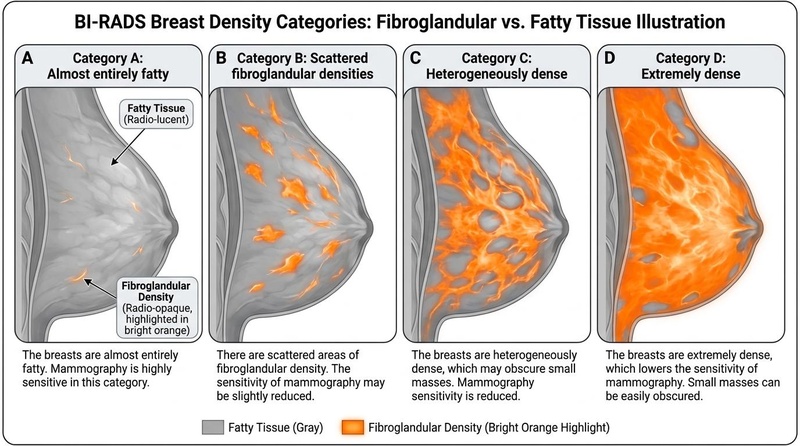
Breast density refers to the relative proportion of fibroglandular tissue to adipose (fatty) tissue within the breast, as assessed on a mammogram [12:1]. Under the Breast Imaging Reporting and Data System (BI-RADS), breast density is classified into categories ranging from non-dense tissue to dense tissue (including heterogeneously dense and extremely dense breasts) [12:2].
- Non-Dense Breasts: Consist of tissue where the sensitivity of digital mammography is high, meaning supplemental screening is not clinically indicated [12:3].
- Dense Breasts: Consist of heterogeneously dense or extremely dense tissue, which limits the sensitivity of mammography, masking lesions and representing an independent risk factor for breast cancer [12:4][11:3].
¶ The Dual Impact of Dense Breasts
- The Masking Effect: Fibroglandular tissue blocks X-rays and appears white on a mammogram, similar to breast cancers. Consequently, dense tissue can obscure or "mask" an underlying malignancy, limiting the detection of cancer with mammography [11:4].
- Independent Risk Factor: Extremely dense breast tissue is an independent risk factor for the development of breast cancer, representing increased risk [11:5].
¶ Screening Protocols & Guidelines
¶ Average-Risk Screening Protocols
Average risk is defined as having a lifetime breast cancer risk of less than 15% (e.g., no personal history, no strong family history, and no known genetic susceptibility). Clinical organizations agree on the utility of screening but maintain divergent guidance regarding the age of initiation, cessation, and optimal screening intervals:
| Organization | Age to Start | Screening Interval | Age to Stop | Modality Recommendation |
|---|---|---|---|---|
| USPSTF (2024) [1:7] | 40 years | Biennial (Every 2 years) | 74 years [1:8] | Digital Mammography or DBT |
| ACS (2015) [9:1] | 45 years | Annual or biennial (noting premenopausal annual benefits) [9:2] | Continuing as long as overall health is good and life expectancy is ≥10 years (supports screening ≥70 in good health) [9:3] | Regular screening mammography |
| ACOG (Clinical Practice Update 2025 / PB 179) [16][17] | 40 years [16:1] | Shared decision-making (various intervals) [16:2] | Based on health status & life expectancy [16:3] | Mammography (standard 2D/3D) |
| ACR/SBI Guidelines (based on Weinstein 2021 & Monticciolo 2024) [12:5][6:2] | 40 years [6:3] | Annual [6:4] | No upper age limit (supports screening up to age 79 based on lifetime benefits) [6:5] | Digital breast tomosynthesis (DBT) or mammography [12:6] |
Guideline recommendations for average-risk individuals show broad consensus on the benefit of screening starting at age 40 or 45, though they differ on the optimal screening interval (annual vs. biennial) and cessation criteria [1:9][9:4][16:4][17:1][12:7][6:6]:
- USPSTF (2024) recommends biennial screening mammography for women aged 40 to 74 years, concluding with moderate certainty that this strategy has a moderate net benefit [1:10].
- ACS (2015): The American Cancer Society (ACS) recommends that women at average risk for breast cancer begin regular screening mammography at age 45 (strong recommendation) [9:5]. Under this guideline, women aged 45 to 54 should be screened annually (qualified recommendation), while those aged 55 and older should transition to biennial screening or have the opportunity to continue screening annually (qualified recommendation) [9:6]. Additionally, women should have the opportunity to begin annual screening between ages 40 and 44 (qualified recommendation), and screening should continue as long as overall health is good and life expectancy is 10 years or longer (qualified recommendation) [9:7].
- ACOG (Practice Bulletin No. 179 / Clinical Practice Update 2025): Under the ACOG guideline framework, regular screening mammography starting at age 40 years is recognized to reduce breast cancer mortality in average-risk women, utilizing a framework of shared decision-making to assist women in choosing screening ages and intervals from within a range of reasonable options [16:5]. ACOG's 2025 Clinical Practice Update provides focused, revised guidance on the age to start routine breast cancer screening with mammography, updating these previous guideline selections [17:2].
- ACR/SBI Guidelines (based on Weinstein 2021 and Monticciolo 2024): In clinical updates, annual breast cancer screening starting at age 40 provides the greatest mortality reduction and gains the most life-years compared to biennial screening or starting at later ages [6:7]. The ACR Appropriateness Criteria note that digital breast tomosynthesis (DBT) improves screening sensitivity in all women, and recommend that average-risk individuals with dense breasts discuss supplemental screening options (such as breast MRI, abbreviated MRI, or ultrasound) based on tissue density and risk [12:8].
¶ High-Risk & Genetic Screening Pathways
High-risk individuals are defined as having an elevated cumulative lifetime risk of breast cancer, typically driven by [16:6][12:9]:
- Known pathogenic germline mutations in high-penetrance genes (e.g., BRCA1, BRCA2).
- A strong family history of breast or ovarian cancer (without genetic testing).
- A history of therapeutic chest radiation or other high-risk factors.
For high-risk cohorts, clinical guidelines recommend intensified, multi-modality screening regimens. Specifically, guidelines from the American College of Radiology (ACR) [12:10] recommend that high-risk individuals—including those with known pathogenic germline mutations (such as BRCA1 or BRCA2), a strong family history of breast cancer, or a history of therapeutic chest radiation—undergo annual supplemental breast MRI in addition to screening mammography [12:11], with the specific timing, age of initiation, and intervals individualized in close consultation with a clinician. The American College of Obstetricians and Gynecologists (ACOG) highlights the importance of early hereditary risk assessment, genetic counseling, and clinical referral to establish tailored, risk-reduction strategies for these patients [18][19].
¶ Screening vs. Surveillance vs. Diagnostic Evaluation
It is vital to distinguish these clinical pathways, as they dictate different protocols and clinical criteria:
- Screening Evaluation: Performed on strictly asymptomatic individuals at standard intervals. It involves standard 4-view imaging (CC and MLO views of both breasts) interpreted by a radiologist, often asynchronously.
- Surveillance Evaluation: Performed on asymptomatic individuals with a personal history of treated breast cancer. Consensus guidelines from the American Society of Clinical Oncology (ASCO), the American Cancer Society (ACS), and the American College of Radiology (ACR) recommend regular surveillance with history, physical exam, and annual mammography or tomosynthesis, while avoiding routine systemic laboratory or advanced imaging tests in asymptomatic patients [20][21].
- Diagnostic Evaluation: Performed on individuals presenting with clinical symptoms or those with abnormal screening findings. Diagnostic evaluations utilize targeted diagnostic imaging (such as diagnostic mammography and/or ultrasound) and potential biopsy (such as core needle biopsy) to characterize clinically suspect masses [22][23].
¶ Clinical Classifications (BI-RADS)
The Breast Imaging Reporting and Data System (BI-RADS) categories are used in clinical practice to classify breast MRI and mammographic findings, ranging from Category 0 (incomplete) to Category 6 (known biopsy-proven malignancy) [12:12]:
| BI-RADS Category | Assessment | Likelihood of Malignancy | Standard Recommended Clinical Action |
|---|---|---|---|
| 0 | Incomplete | N/A | Needs additional diagnostic imaging (mammographic views or ultrasound) and/or comparison with prior exams [12:13]. |
| 1 | Negative | Negative / Normal findings | Continue routine screening at standard risk-stratified intervals [12:14]. |
| 2 | Benign Findings | Benign (non-malignant) | Continue routine screening. Finding is non-malignant (e.g., simple cysts, benign calcifications) [12:15]. |
| 3 | Probably Benign | Probably benign | Short-interval follow-up imaging (typically 6 months) to establish stability. Biopsy is optional but rarely indicated [12:16]. |
| 4 | Suspicious | Suspicious finding | Tissue biopsy is required. Classifications include: 4A (low suspicion), 4B (moderate), and 4C (high) [12:17]. |
| 5 | Highly Suggestive of Malignancy | Highly suggestive | Immediate tissue biopsy and surgical/oncological consultation [12:18]. |
| 6 | Known Biopsy-Proven Malignancy | Known malignancy | Action dictated by clinical staging and therapeutic plan (typically surgical resection, chemotherapy, or radiation) [12:19]. |
¶ Evidence on Screening Outcomes
¶ Landmark Clinical Trials
The efficacy of breast cancer screening is established through several multi-decade randomized controlled trials:
- The Swedish Two-County Trial: Initiated in 1977, this trial randomized 133,065 women to screening invitation or usual care. The 29-year follow-up confirmed a highly significant reduction in breast cancer-specific mortality in the screened group, with a relative risk [RR] of 0.69 (by local end point committee data) or 0.73 (by consensus data), as evaluated in systematic reviews [24]. Taking cluster randomization into account, analysis of the trial data confirmed a significant 30% reduction in breast cancer mortality among the screened population [25]. The trial demonstrated that the absolute benefit of screening continues to expand beyond 20 years of follow-up.
- The Malmo Mammographic Screening Trial: The Malmo Mammographic Screening Trial was part of the Swedish randomized trials overview, which reported 511 breast cancer deaths over 1,864,770 person-years in the invited groups versus 584 breast cancer deaths over 1,688,440 person-years in the control groups, representing a significant 21% reduction in breast cancer mortality (RR = 0.79, 95% CI: 0.70-0.89), as evaluated in systematic reviews [3:3]. Furthermore, the long-term relative reduction has been confirmed after more than 20 years of follow-up in the Swedish randomized controlled mammography trials in Malmo, Stockholm, and Goteborg [5:1].
¶ CISNET Modeling Evidence
Because large RCTs are expensive and take decades to complete, the National Cancer Institute's Cancer Intervention and Surveillance Modeling Network (CISNET) utilizes comparative simulation modeling to evaluate various screening scenarios.
The CISNET 2023 modeling estimates demonstrated that [6:8]:
- Annual screening from ages 40 to 79 improved breast cancer mortality reduction compared with biennial screening from ages 50 to 74 and biennial screening from ages 40 to 74, yielding mortality reduction percentages of 41.7%, 25.4%, and 30.0%, respectively.
- The annual screening strategy from ages 40 to 79 averted the most breast cancer deaths and gained the most life-years compared to other analyzed screening scenarios.
¶ Evidence summary table (human outcomes)
| Outcome / Goal | Effect* | Consistency | Evidence quality | Trials | Notes (population, duration, dose) |
|---|---|---|---|---|---|
| Breast Cancer Mortality Reduction | High | High | Multiple RCTs | 15% to 21% reduction in clinical trials [5:2][3:4], up to 41.7% reduction in simulation models [6:9] for screening starting at age 40. | |
| All-Cause Mortality | High | High | Multiple RCTs | Furthermore, systematic reviews of clinical trials have not demonstrated a statistically significant reduction in all-cause mortality [4:2][3:5]. This is expected, as breast cancer-specific deaths represent only a small fraction of overall deaths in these cohorts, making trials under-designed to detect an overall mortality difference. | |
| False Positive Recalls | High | High | Observational Cohorts | Lifetime cumulative risk of false-positive results is greater with younger starting ages and annual screening intervals [9:8][1:11]. | |
| Benign Biopsies | High | High | Observational Cohorts | Although annual screening may detect cancers earlier, standard guidelines indicate it significantly increases harms, including cumulative false-positives and biopsies compared to biennial screening [1:12]. | |
| Overdiagnosis / Overtreatment | Moderate | Moderate | Observational Cohorts | Significant risk of overdiagnosis and overtreatment shown in reviews and modeling, estimating 10% or less in observational studies [7:1], 11% to 19% in trials [26], and up to 30% in Cochrane overviews [4:3]. | |
| Radiation-Induced Cancer Risk | High | High | Modeling | Screening mammography can be performed at acceptable levels of radiation risk, with clinical mortality benefits offsetting radiation concerns [1:13][2:6][6:10]. |
*Effect column is based on the compact renderer: e="[dir][mag][impact]" where dir = u (up), d (down), e (equal), q (unclear); mag = 0 to 3 (magnitude); and impact = p (positive), n (negative), x (neutral/unknown).
¶ Safety, Harms, and Downstream Cascades
¶ False Positives & False Negatives
- False Positives: A false-positive result occurs when a screening test is flagged as abnormal but subsequent diagnostic workup reveals no malignancy. While the recall rate for a single screening round is low (ranging from 6.5% to 9.6% depending on the scenario) [6:11], the cumulative lifetime risk of false-positive examination results is significantly greater when screening begins at younger ages (due to a greater number of lifetime mammograms) and with more frequent (annual) screening intervals [9:9][1:14].
- False Negatives: A false-negative result occurs when a cancer is missed during screening. The masking effect of dense breasts significantly reduces the sensitivity of standard mammography, obscuring underlying malignancies and increasing the clinical risk of missed or delayed diagnoses [11:6][2:7].
¶ Overdiagnosis & Overtreatment
Overdiagnosis is defined as the detection of true histological malignancies (including ductal carcinoma in situ [DCIS] and slow-growing invasive adenocarcinomas) that would never have progressed to cause clinical symptoms or death in the patient’s lifetime.
- Mechanism: Because it is currently impossible to distinguish progressive from non-progressive lesions at the time of biopsy, standard practice dictates treating all detected cancers with surgical resection, radiation, and/or systemic endocrine therapy.
- Rates: Estimates of overdiagnosis vary depending on study design and methodology. Contemporary systematic reviews and modeling suggest that the overdiagnosis rate ranges from 11% to 19% in some clinical trial analyses [26:1], up to 30% in Cochrane overviews [4:4], while recent registry-based observational studies suggest it is 10% or less [7:2].
¶ Downstream Procedures & Psychological Harms
A positive screening mammogram triggers a cascade of downstream clinical interventions:
- Diagnostic Mammography & Targeted Ultrasound: High-resolution views to characterize the lesion [22:1][23:1].
- Core Needle Biopsy: Performed under stereotactic, tomosynthesis, or ultrasound guidance to obtain tissue [22:2][23:2].
- Overtreatment & Physical Harms: Routine screening increases the rates of treatment for lesions that represent overdiagnosis, leading to unnecessary therapeutic interventions in otherwise healthy individuals [2:8][7:3].
¶ Radiation Safety & Exposure
Mammography exposes patients to low-dose ionizing radiation. Comprehensive clinical evidence indicates that screening mammography can be performed at acceptable levels of radiation risk, with the long-term mortality reduction benefits substantially offsetting radiation concerns [1:15][2:9][6:12].
¶ Shared Decision-Making & Stopping Considerations
¶ Risk-Harm Trade-offs & Personal Values
While some guidelines recommend routine screening starting at age 40, clinical guidelines from ACOG and other major panels emphasize that shared decision-making is essential to assist patients in balancing personal values regarding screening benefits against potential harms [16:7][8:1]. This discussion is particularly relevant because starting screening at a younger age (such as during the 40s) increases the cumulative lifetime risk of false-positive results and benign biopsies [9:10][16:8]. Patient decision aids are designed to make the decision explicit, provide evidence-based information about options, benefits, and harms, and help clarify personal values to support informed decision-making [8:2]. Clinicians and patients should evaluate:
- Personal Risk Profile: Calculated via tools like the Tyrer-Cuzick or Gail model [27].
- Breast Density Status: Knowing if Category C/D density warrants supplemental imaging [11:7][12:20].
- Value Preferences: Some individuals prioritize maximizing cancer detection (accepting a higher risk of false positives and biopsies), while others prioritize avoiding diagnostic cascades and overdiagnosis (preferring biennial intervals or starting screening at a later age) [8:3].
¶ Stopping Screening: Chronological Age vs. Biological Life Expectancy
Divergent recommendations exist regarding when to stop routine screening:
- The Chronological Approach: The USPSTF 2024 guidelines conclude that the evidence is insufficient to assess the balance of benefits and harms in women aged 75 or older [1:16]. This is driven by the lack of randomized clinical trial data for this cohort.
- The Biological/Life Expectancy Approach: The ACS recommends continuing regular screening as long as the patient's overall health is good and their estimated life expectancy is at least 10 years [9:11]. This reflects the clinical reality that the benefits of early detection require approximately 5 to 10 years of lag time to translate into a survival benefit [9:12].
¶ Clinical Red Flags (Symptomatic Presentation)
Breast cancer screening is strictly designed for asymptomatic individuals. If any of the following clinical "red flags" are present, standard screening protocols are inappropriate, and a comprehensive diagnostic evaluation is immediately indicated:
- Palpable Breast Mass: Any new, distinct palpable mass or focal lump in the breast or axilla requires systematic diagnostic evaluation. The masking effect of dense breast tissue significantly reduces the sensitivity of mammography, which can obscure small cancers [11:8][12:21]. Clinical guidelines emphasize that because mammography has lower sensitivity in dense tissue, a negative screening mammogram should not delay further diagnostic evaluation—including targeted ultrasound or biopsy—if there is a clinically suspicious palpable mass [1:17][12:22][16:9]. Under these scenarios, diagnostic procedures—including further imaging techniques and fine-needle aspiration or core biopsy—should be utilized before resorting to surgical biopsy [22:3].
- New Unilateral Nipple Retraction: A new, persistent pulling inward or inversion of one nipple.
- Spontaneous Nipple Discharge: Unilateral discharge from a single duct, particularly if it is bloody, serous (clear/yellow), or occurs without manual expression.
- Skin Changes: Dimpling of the skin resembling an orange peel (peau d'orange), localized redness (erythema), scaling, thickening, or ulceration of the nipple or breast skin.
- Localized Breast Pain: Persistent, non-cyclical, unilateral breast pain focused in a specific location (though pain is rarely the sole presenting symptom of breast cancer).
- Lymphadenopathy: Hard, fixed, or enlarging lymph nodes in the axilla, infraclavicular, or supravicular regions.
¶ Clinician Discussion Checklist
Patients should utilize this clinical checklist to guide their shared decision-making discussion with their primary care physician or gynecologist:
- What is my calculated lifetime breast cancer risk? (Ask to run a validated tool like the Tyrer-Cuzick model).
- What is my breast density category from my most recent mammogram? (Confirm if it is Category C or D).
- Given my density and risk profile, should I consider supplemental screening with Abbreviated Breast MRI or Ultrasound?
- What screening interval (annual vs. biennial) is most appropriate for my risk profile and personal preferences?
- At what age is it appropriate for me to begin screening, and what criteria will we use to determine when to stop?
- What is our clinical plan if my screening mammogram returns an abnormal result (BI-RADS 0 or 3)?
¶ FAQ
¶ Is 3D mammography (DBT) truly superior to 2D mammography?
Clinical trials and large-scale registry data show that 3D tomosynthesis (DBT) detects more invasive breast cancers per 1000 screens at the first screening round than digital mammography (DM) alone [2:10]. While some guidelines (such as the ACR/SBI Guidelines) highlight DBT's improved sensitivity [12:23], other guidelines (such as the USPSTF 2024) recommend digital mammography or DBT without expressing a formal preference, noting that the long-term relative benefits remain under study [1:18][2:11].
¶ Can I skip mammograms and just do breast ultrasounds?
No. Ultrasound is not validated as a standalone screening tool for average-risk individuals. It is associated with a high rate of false-positive findings and additional biopsies [2:12]. It should only be used as an adjunct to mammography in patients with dense breasts, as evaluated in systematic reviews [10:1][12:24].
¶ What is an abbreviated breast MRI (Ab-MRI)?
An abbreviated breast MRI is a shortened version of a standard breast MRI. It focuses strictly on the contrast-enhanced dynamic sequences required to identify highly vascular tumor tissues. Clinical studies indicate that abbreviated breast MRI screening represents a potential method to reduce screening workloads, costs, and scan times while maintaining high diagnostic accuracy comparable to full protocols [14:2].
¶ Why did the USPSTF change its starting age recommendation from 50 to 40 in 2024?
The USPSTF recommends biennial screening mammography starting at age 40 instead of 50, concluding with moderate certainty that biennial screening in women aged 40 to 74 years has a moderate net benefit [1:19]. This is supported by comparative modeling estimates (CISNET) projecting that screening starting at age 40 improves overall breast cancer mortality reduction compared to starting at age 50 [6:13].
¶ If I have dense breasts (BI-RADS C or D), what is my optimal screening regimen?
If you have dense breasts, guidelines suggest discussing supplemental screening options based on tissue density and risk [12:25]. While the USPSTF (2024) average-risk screening recommendations remain biennial (every 2 years) and conclude that evidence is insufficient to recommend supplemental screening regardless of density [1:20], other guidelines and evidence-based reviews indicate that intermediate-risk individuals with dense breasts may benefit from supplemental contrast-enhanced mammography, screening ultrasound, or breast MRI [10:2][12:26]. For extremely dense breasts (Category D), the addition of supplemental Breast MRI is associated with a highly significant reduction in interval cancers [11:9].
¶ References
US Preventive Services Task Force, Nicholson WK, Silverstein M, et al. Screening for Breast Cancer: US Preventive Services Task Force Recommendation Statement. JAMA. 2024;331(22):1918-1930. https://pubmed.ncbi.nlm.nih.gov/38687503/ ↩︎ ↩︎ ↩︎ ↩︎ ↩︎ ↩︎ ↩︎ ↩︎ ↩︎ ↩︎ ↩︎ ↩︎ ↩︎ ↩︎ ↩︎ ↩︎ ↩︎ ↩︎ ↩︎ ↩︎ ↩︎
Henderson JT, Webber EM, Weyrich MS. Screening for Breast Cancer: Evidence Report and Systematic Review for the US Preventive Services Task Force. JAMA. 2024;331(22):1931-1943. https://pubmed.ncbi.nlm.nih.gov/38687490/ ↩︎ ↩︎ ↩︎ ↩︎ ↩︎ ↩︎ ↩︎ ↩︎ ↩︎ ↩︎ ↩︎ ↩︎ ↩︎
Nystrom L, Andersson I, Bjurstam N, et al. Long-term effects of mammography screening: updated overview of the Swedish randomised trials. Lancet. 2002;359(9310):909-914. https://pubmed.ncbi.nlm.nih.gov/11918907/ ↩︎ ↩︎ ↩︎ ↩︎ ↩︎ ↩︎
Gotzsche PC, Jorgensen KJ. Screening for breast cancer with mammography. Cochrane Database of Systematic Reviews. 2013. https://pubmed.ncbi.nlm.nih.gov/23737396/ ↩︎ ↩︎ ↩︎ ↩︎ ↩︎
Nystrom L, Bjurstam N, Jonsson H, et al. Reduced breast cancer mortality after 20+ years of follow-up in the Swedish randomized controlled mammography trials in Malmo, Stockholm, and Goteborg. Journal of Medical Screening. 2017;24(1):34-42. https://pubmed.ncbi.nlm.nih.gov/27306511/ ↩︎ ↩︎ ↩︎
Monticciolo DL, Hendrick RE, Helvie MA. Outcomes of Breast Cancer Screening Strategies Based on Cancer Intervention and Surveillance Modeling Network Estimates. Radiology. 2024;250(2):e231179. https://pubmed.ncbi.nlm.nih.gov/38376405/ ↩︎ ↩︎ ↩︎ ↩︎ ↩︎ ↩︎ ↩︎ ↩︎ ↩︎ ↩︎ ↩︎ ↩︎ ↩︎ ↩︎
Hendrick RE, Monticciolo DL. USPSTF Recommendations and Overdiagnosis. Journal of Breast Imaging. 2024. https://pubmed.ncbi.nlm.nih.gov/38865364/ ↩︎ ↩︎ ↩︎ ↩︎
Stacey D, Lewis KB, Smith M, et al. Decision aids for people facing health treatment or screening decisions. Cochrane Database of Systematic Reviews. 2024. https://pubmed.ncbi.nlm.nih.gov/38284415/ ↩︎ ↩︎ ↩︎ ↩︎
Oeffinger KC, Fontham ET, Etzioni R, et al. Breast Cancer Screening for Women at Average Risk: 2015 Guideline Update From the American Cancer Society. JAMA. 2015;314(16):1599-1614. https://pubmed.ncbi.nlm.nih.gov/26501536/ ↩︎ ↩︎ ↩︎ ↩︎ ↩︎ ↩︎ ↩︎ ↩︎ ↩︎ ↩︎ ↩︎ ↩︎ ↩︎
Hussein H, Abbas E, Keshavarzi S, et al. Supplemental Breast Cancer Screening in Women with Dense Breasts and Negative Mammography: A Systematic Review and Meta-Analysis. Radiology. 2023;306(3):e221788. https://pubmed.ncbi.nlm.nih.gov/36719288/ ↩︎ ↩︎ ↩︎
Bakker MF, de Lange SV, Pijnappel RM, et al. Supplemental MRI Screening for Women with Extremely Dense Breast Tissue. New England Journal of Medicine. 2019;381(22):2091-2102. https://pubmed.ncbi.nlm.nih.gov/31774954/ ↩︎ ↩︎ ↩︎ ↩︎ ↩︎ ↩︎ ↩︎ ↩︎ ↩︎ ↩︎
Expert Panel on Breast Imaging, Weinstein SP, Slanetz PJ, et al. ACR Appropriateness Criteria® Supplemental Breast Cancer Screening Based on Breast Density. Journal of the American College of Radiology. 2021;18(11):S456-S473. https://pubmed.ncbi.nlm.nih.gov/34794600/ ↩︎ ↩︎ ↩︎ ↩︎ ↩︎ ↩︎ ↩︎ ↩︎ ↩︎ ↩︎ ↩︎ ↩︎ ↩︎ ↩︎ ↩︎ ↩︎ ↩︎ ↩︎ ↩︎ ↩︎ ↩︎ ↩︎ ↩︎ ↩︎ ↩︎ ↩︎ ↩︎
Veenhuizen SGA, de Lange SV, Bakker MF, et al. Supplemental Breast MRI for Women with Extremely Dense Breasts: Results of the Second Screening Round of the DENSE Trial. Radiology. 2021;299(1):73-83. https://pubmed.ncbi.nlm.nih.gov/33724062/ ↩︎
van Grinsven SEL, Mann RM, Monninkhof EM, et al. Multireader Diagnostic Accuracy of Abbreviated Breast MRI for Screening Women with Extremely Dense Breasts. Radiology. 2025. https://pubmed.ncbi.nlm.nih.gov/40392091/ ↩︎ ↩︎ ↩︎
Gilbert FJ, Payne NR, Allajbeu I, et al. Comparison of supplemental breast cancer imaging techniques-interim results from the BRAID randomised controlled trial. Lancet. 2025. https://pubmed.ncbi.nlm.nih.gov/40412427/ ↩︎
American College of Obstetricians and Gynecologists. Practice Bulletin Number 179: Breast Cancer Risk Assessment and Screening in Average-Risk Women. Obstetrics & Gynecology. 2017;130(1):e1-e16. https://pubmed.ncbi.nlm.nih.gov/28644335/ ↩︎ ↩︎ ↩︎ ↩︎ ↩︎ ↩︎ ↩︎ ↩︎ ↩︎ ↩︎
American College of Obstetricians and Gynecologists. Age to Initiate Routine Breast Cancer Screening: ACOG Clinical Practice Update. Obstetrics & Gynecology. 2025. https://pubmed.ncbi.nlm.nih.gov/39388713/ ↩︎ ↩︎ ↩︎
American College of Obstetricians and Gynecologists. ACOG Practice Bulletin No. 103: Hereditary breast and ovarian cancer syndrome. Obstetrics & Gynecology. 2009;113(4):957-966. https://pubmed.ncbi.nlm.nih.gov/19305347/ ↩︎
American College of Obstetricians and Gynecologists. Hereditary Cancer Syndromes and Risk Assessment: ACOG COMMITTEE OPINION, Number 793. Obstetrics & Gynecology. 2019;134(6):e143-e190. https://pubmed.ncbi.nlm.nih.gov/31764758/ ↩︎
Mehta TS, Lourenco AP, Niell BL, et al. ACR Appropriateness Criteria® Imaging After Breast Surgery. Journal of the American College of Radiology. 2022;19(11):S337-S353. https://pubmed.ncbi.nlm.nih.gov/36436961/ ↩︎
Runowicz CD, Leach CR, Henry NL, et al. American Cancer Society/American Society of Clinical Oncology Breast Cancer Survivorship Care Guideline. CA: A Cancer Journal for Clinicians. 2016;66(1):43-73. https://pubmed.ncbi.nlm.nih.gov/26641959/ ↩︎
Tabar L, Fagerberg G, Duffy SW. Update of the Swedish two-county program of mammographic screening for breast cancer. Radiologic Clinics of North America. 1992;30(1):187-210. https://pubmed.ncbi.nlm.nih.gov/1732926/ ↩︎ ↩︎ ↩︎ ↩︎
Bevers TB, Helvie M, Bonaccio E, et al. Breast Cancer Screening and Diagnosis, Version 3.2018, NCCN Clinical Practice Guidelines in Oncology. Journal of the National Comprehensive Cancer Network. 2018;16(11):1362-1389. https://pubmed.ncbi.nlm.nih.gov/30442736/ ↩︎ ↩︎ ↩︎
Tabar L, Vitak B, Chen TH, et al. Swedish two-county trial: impact of mammographic screening on breast cancer mortality during 3 decades. Radiology. 2011;260(3):658-663. https://pubmed.ncbi.nlm.nih.gov/21712474/ ↩︎
Duffy SW, Tabar L, Vitak B, et al. The Swedish Two-County Trial of mammographic screening: cluster randomisation and end point evaluation. Annals of Oncology. 2003;14(8):1196-1202. https://pubmed.ncbi.nlm.nih.gov/12881376/ ↩︎
Independent UK Panel on Breast Cancer Screening. The benefits and harms of breast cancer screening: an independent review. The Lancet. 2012;380(9855):1778-1786. https://pubmed.ncbi.nlm.nih.gov/23117178/ ↩︎ ↩︎
Liu L, Zhou P, Hou L, et al. Development and performance of female breast cancer incidence risk prediction models: a systematic review and meta-analysis. Annals of Medicine. 2025. https://pubmed.ncbi.nlm.nih.gov/40684451/ ↩︎