| Primary Function | Restoration, Repair, Regulation |
| Optimal Duration | 7-8 hours/night (adults) |
| Key Metrics | Duration, Quality, Regularity, Latency |
| Core Mechanism | Glymphatic Clearance, Autophagy, Molecular Clocks |
| Impacts | Metabolic, Immune, Cognitive, Cardiovascular |
| Accessibility | Universal, Low Cost (Lifestyle) |
¶ Sleep
Sleep is a fundamental physiological process critical for longevity, health maintenance, and optimal aging. It enables cellular repair, metabolic regulation, and brain waste clearance[1][2]. Across aging, sleep quality becomes less efficient and regulatory mechanisms deteriorate, contributing to accelerated biological aging and increased disease risk[3][4].
¶ At a glance
¶ Key Points
- Day-to-day regularity is a stronger predictor of longevity than sleep duration alone; consistent timing reduces all-cause mortality by up to 48%[5].
- Optimal duration falls between 7–8 hours for most adults; both short (<7h) and long (>9h) sleep show U-shaped correlations with increased mortality, though long sleep is often confounded by underlying illness[6][7].
- Glymphatic clearance occurs primarily during slow-wave (deep) sleep, clearing neurotoxic proteins like amyloid-beta and tau[2:1][8].
- Critical health marker: Poor sleep and circadian disruption are linked to a 28% increased risk of type 2 diabetes and profound cardiovascular dysfunction[9][10].
¶ What People Use It For
- Primary Goal: Lifespan and healthspan extension, neurological waste clearance, hormone optimization, cardiovascular health, and immune system resilience.
- Evidence Quality (Overall): High-quality evidence based on massive prospective cohorts and clinical guidelines.
¶ Bottom Line
Optimizing sleep via consistency, environment, and circadian alignment represents the highest-leverage, lowest-cost non-pharmacological lifestyle intervention available to reduce biological age and delay chronic disease onset.
CRITICAL CLINICAL CRITERIA
Severe Obstructive Sleep Apnea (OSA) carries a 2.25-fold increased risk of cardiovascular death if left untreated; immediate clinical assessment and continuous positive airway pressure (CPAP) therapy are required for clinical resolution[11][12].
Chronic Benzodiazepine/Z-Drug Use for insomnia carries significant safety risks including falls, cognitive decline, and addiction in older adults, with minimal long-term therapeutic benefit compared to CBT-I[13][14].
Neurological Parasomnias: Dream-enactment behaviors during REM sleep are strong prodromal indicators of alpha-synucleinopathies, carrying up to an 80% risk of conversion to Parkinson's disease or Lewy body dementia within 10-15 years[15].
Sleep Optimization Protocol
To systematically optimize sleep architecture, regularity, and restorative efficiency, implement the following tiered protocol:
¶ Tier 1: The Circadian & Regularity Baseline
- Set an Anchor Wake Time: Wake up at the exact same time every day (including weekends) within a ±30-minute window[5:1].
- Early Light Exposure: View 10–15 minutes of bright, direct sunlight within 1 hour of waking to anchor the circadian clock and suppress melatonin[16].
- Optimized Sleep Environment: Maintain a bedroom temperature of 60–67°F (15–19°C), complete blackout (using eye masks or blackout shades), and minimize ambient noise (<30 dB)[17].
¶ Tier 2: The Evening Wind-Down & Light Hygiene
- The 3-2-1 Rule:
- 3 hours before bed: Stop eating large meals and consuming alcohol (which disrupts REM sleep and triggers late-night fragmentation)[17:1][18].
- 2 hours before bed: Cease heavy working and cognitive stressors.
- 1 hour before bed: Turn off all bright screens (phones, TVs, computers) or use strict red/amber lighting to prevent melatonin suppression[19].
- Late-Day Stimulant Block: Avoid caffeine intake within 10 hours of bed (due to its ~5-hour half-life and adenosine receptor blocking effects)[17:2][20].
¶ Tier 3: Evidence-Based Supplemental Stack (Take 30–60 minutes pre-bed)
For a comprehensive clinical overview of sleep-promoting ingredients and combinations, see the dedicated Sleep Starter Kit Stack.
- Magnesium Bisglycinate: 200–400 mg (improves GABAergic signaling and somatic relaxation)[21].
- Glycine: 3 grams (lowers core body temperature, decreasing sleep latency and improving deep sleep quality)[22][23].
- L-Theanine: 100–200 mg (increases alpha-wave brain activity and reduces sleep-onset latency)[24].
¶ What is Sleep?
Sleep is a highly active, regulated physiological state essential for systemic homeostasis. Far from passive rest, sleep is characterized by complex neurological transitions, somatic repair cycles, and metabolic transformations. It is defined behaviorally by reduced responsiveness to external stimuli, stereotyped postures, and rapid reversibility (distinguishing it from coma or anesthesia)[25]. Sleep is divided into two distinct states: Non-Rapid Eye Movement (NREM) sleep (further subdivided into light N1/N2 and slow-wave N3 deep sleep) and Rapid Eye Movement (REM) sleep, each governed by specific subcortical networks and chemical neurotransmitters[25:1][26].
¶ Core Regulators: The Two-Process Model
Sleep regulation is dictated by the interaction of two distinct biological forces: the circadian clock (Process C) and the homeostatic sleep drive (Process S).
WAKING: Adenosine Accumulation (Process S Peaks)
|=========================================> Bedtime (Sleep pressure high)
LIGHT: Entrains SCN (Process C Suppresses Melatonin)
|-----------------------------------------> Sunset (Melatonin rises)
¶ Process C: Circadian Rhythm Biology
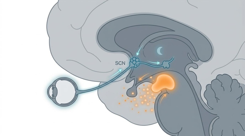
Process C is the endogenous, ~24-hour rhythmic oscillator driven by the master pacemaker: the suprachiasmatic nucleus (SCN), situated in the anterior hypothalamus[27]. The SCN coordinates timing across the entire organism by entraining peripheral tissue clocks located in the liver, skeletal muscle, gut, and cardiovascular tissues to match environmental light-dark cycles[28].
At the cellular level, the circadian rhythm is driven by an autonomous, molecular transcription-translation feedback loop (TTFL) in virtually all cells:
- CLOCK and BMAL1 heterodimerize in the nucleus and bind to E-box elements to drive transcription of Period (PER1, PER2, PER3) and Cryptochrome (CRY1, CRY2) genes[29].
- PER and CRY proteins accumulate in the cytoplasm, homodimerize, and translocate back into the nucleus.
- Once in the nucleus, PER-CRY complexes physically interact with the CLOCK-BMAL1 heterodimer, inhibiting its transcriptional activity[30][29:1].
- Over the course of the day, PER and CRY proteins are progressively phosphorylated, targeted for ubiquitination, and degraded by the proteasome, releasing CLOCK-BMAL1 from inhibition and starting the 24-hour cycle anew[30:1].
The SCN is primarily entrained by light. Specialized intrinsically photosensitive retinal ganglion cells (ipRGCs) in the retina project directly to the SCN via the retinohypothalamic tract[31]. These ipRGCs express the photopigment melanopsin, which is highly sensitive to short-wavelength blue light (460–480 nm)[31:1]. Activation of ipRGCs signals the SCN to suppress pineal melatonin synthesis during daylight hours.
¶ Process S: Sleep Pressure & Adenosine
Process S represents the homeostatic sleep drive—the accumulating neurochemical pressure to sleep that builds linearly during wakefulness and dissipates during sleep[3:1].
The primary mediator of Process S is adenosine, a purine nucleoside generated as a byproduct of metabolic activity. During wakefulness, high-energy adenosine triphosphate (ATP) is consumed by active brain cells to fuel physiological processes. This intracellular ATP cleavage yields adenosine, which is progressively transported into the extracellular space of the basal forebrain and cerebral cortex[32].
Extracellular adenosine binds to two primary receptor subtypes in sleep-regulatory centers:
- Adenosine Receptors: Inhibitory G-protein-coupled receptors. Binding of adenosine to receptors inhibits the activity of wake-promoting neurotransmitter systems (such as cholinergic neurons in the basal forebrain and histaminergic neurons in the tuberomammillary nucleus)[6:1].
- Adenosine Receptors: Excitatory G-protein-coupled receptors. Binding of adenosine to receptors excites GABAergic neurons in the ventrolateral preoptic nucleus (VLPO), which in turn project to and inhibit wake-promoting centers, initiating NREM sleep[6:2].
During sleep, metabolic demand drops, and astrocytic and enzymatic clearance mechanisms actively deplete extracellular adenosine, resetting the homeostatic sleep drive to baseline. Caffeine acts as a competitive antagonist of both and receptors, blocking the binding of endogenous adenosine and masking the sensation of sleep pressure without actually clearing the accumulated nucleoside[20:1].
¶ Main Benefits
¶ Cellular Repair and Genome Protection
During waking hours, metabolic activity and environmental exposures cause cellular wear and accumulate DNA double-strand breaks in neurons[33]. Sleep serves as the primary homeostatic window where DNA damage response pathways (mediated by PARP1, Rad52, Ku80) repair genomic lesions and maintain chromosomal stability[34][32:1]. Simultaneously, sleep triggers autophagy and proteasomal degradation to recycle damaged organelles and prevent proteotoxicity[35][36].
¶ Cognitive Sparing and Waste Clearance
The brain lacks a traditional lymphatic system. During slow-wave sleep, the interstitial space expands by 60%, allowing cerebrospinal fluid to mix with interstitial fluid and flush out metabolic waste, including beta-amyloid and tau proteins[2:2][8:1]. This nocturnal clearance is a critical defense mechanism against neurodegenerative diseases like Alzheimer’s[37].
¶ Metabolic and Hormonal Equilibrium
Sleep regulates the balance of endocrine signals governing energy homeostasis. Adequate sleep maintains normal sensitivity of peripheral tissues to insulin and supports optimal ratios of satiety (leptin) and hunger (ghrelin) hormones[38][39]. Furthermore, slow-wave sleep is the primary driver of pulsatile growth hormone release, which is critical for tissue regeneration and physiological maintenance[40].
¶ Reality Check & Context
¶ Human Reality vs. Animal Hopes
While the broad stroke of sleep science is heavily validated in human clinical cohorts, several precise molecular mechanisms—such as the neuronal chromosome dynamics regulating DNA repair and the precise mathematical models of glymphatic clearance—were discovered in animal models (such as zebrafish and mice)[8:2][34:1]. Direct real-time human visualization of these micro-cellular pathways remains limited due to the invasive nature of current imaging techniques. Nonetheless, human neuroimaging (using functional MRI and CSF flow tracers) has confirmed that slow-wave sleep in humans is indeed accompanied by rhythmic, large-amplitude waves of CSF flow[41], and one night of human sleep deprivation shows detectable increases in amyloid-beta accumulation in vivo[42].
¶ The Commercial Context
The massive rise in consumer sleep trackers (e.g., Oura Ring, Apple Watch, WHOOP, Fitbit) has successfully democratized sleep tracking, shifting user focus toward objective sleep metrics. Validation studies against gold-standard polysomnography show that modern consumer devices are highly sensitive (≥95%) at detecting sleep vs. wake states and tracking total duration[43][44]. However, users should maintain realistic expectations: consumer wearables are significantly less accurate at discriminating specific sleep stages (N1, N2, N3, REM), with sensitivity ranging from 50% to 86%[45]. Furthermore, photoplethysmography (optical) sensors are known to exhibit lower accuracy in individuals with darker skin tones[46]. These tools should be used to track longitudinal trends and consistency, rather than as clinical diagnostics.
¶ Sleep Architecture & Physiology
¶ The Sleep Cycle
Human sleep is organized into structured, cyclic progressions through distinct physiological phases. A standard nocturnal period contains 4 to 5 complete cycles, each spanning 90 to 110 minutes[25:2].
Wakefulness ---> N1 (Light Transition) ---> N2 (Light Sleep / Spindles)
                                              |
REM (Dreams / Paralysis) <--- N3 (Deep Slow-Wave Sleep / Restoration)
- N1 (Light NREM Sleep): The brief transition phase between wakefulness and sleep, marked by a shift from high-frequency beta/alpha waves to theta waves. Somatic responsiveness declines.
- N2 (Light NREM Sleep): Comprising the largest proportion of total sleep time, N2 is characterized electroencephalographically by sleep spindles (bursts of 11–16 Hz activity) and K-complexes (large negative/positive deflections)[25:3]. These features are crucial for sensory gating, memory consolidation, and protecting sleep against external noise.
- N3 (Slow-Wave / Deep Sleep): Characterized by high-amplitude, low-frequency delta waves (0.5–2 Hz). This is the highly restorative phase where physical repair, growth hormone release, and glymphatic clearance occur[8:3][40:1]. N3 dominates the first half of the sleep period.
- REM (Rapid Eye Movement Sleep): A highly active neurological state characterized by rapid saccadic eye movements, low-amplitude mixed-frequency EEG (resembling wakefulness), and profound skeletal muscle atonia (paralysis)[26:1]. REM sleep is critical for emotional processing, neural plasticity, and complex memory integration, and dominates the latter half of the nocturnal period.
¶ Thermoregulation and Sleep
Thermoregulatory biology is deeply intertwined with sleep architecture and sleep-onset pathways[47]. Core body temperature follows a robust circadian rhythm, peaking in the late afternoon and dropping to its lowest point (nadir) roughly 2 hours before habitual wake time[47:1].
The initiation of sleep is triggered by a rapid drop in core body temperature, mediated by a distal-to-proximal skin temperature gradient (DPG)[47:2]. As bedtime approaches, the preoptic area of the hypothalamus signals blood vessels in the hands and feet to dilate. This distal vasodilation promotes heat dissipation to the environment, rapidly cooling the core. Maintaining an ambient room temperature of 60–67°F (15–19°C) prevents peripheral vasoconstriction and accelerates this core-cooling cascade, decreasing sleep-onset latency[17:3]. Conversely, taking a warm bath or shower 90 minutes pre-bed triggers rebound peripheral vasodilation, facilitating a faster rate of subsequent core body cooling[23:1][47:3].
¶ Sex Differences & Hormonal Regulation
Sleep architecture, circadian timing, and susceptibility to sleep disorders display pronounced sex-specific differences, governed primarily by ovarian steroids[48].
- Baseline Architecture: Women exhibit longer self-reported sleep duration, earlier circadian sleep phase, and a higher percentage of slow-wave sleep compared to age-matched men[48:1]. Despite these objective advantages, women report higher subjective sleep disruption and a greater prevalence of chronic insomnia[48:2].
- Hormonal Pacemakers: Progesterone acts as a powerful allosteric modulator of receptors in the central nervous system, exerting anxiolytic, sedative, and muscle-relaxing effects[48:3]. Estrogen coordinates circadian clock genes, facilitates core body temperature drop at night, and preserves healthy airway muscle tone.
- The Menopausal Transition: During perimenopause and menopause, the abrupt decline in progesterone and estrogen disrupts central thermoregulatory networks in the hypothalamus. This disruption triggers vasomotor symptoms (hot flashes and night sweats), which cause severe sleep fragmentation and nocturnal panic[48:4]. Furthermore, post-menopausal women lose the protective upper-airway muscle tone provided by estrogen, resulting in a dramatic surge in Obstructive Sleep Apnea (OSA) risk, approaching rates seen in men[48:5][49].
¶ Modern Sleep Disruptors
¶ Screen Light and Blue Wavelengths
Modern exposure to artificial light at night (ALAN) represents a major threat to circadian alignment. Blue light wavelengths (460–480 nm) stimulate ipRGCs with maximum efficiency, triggering immediate SCN activation and suppressing pineal melatonin synthesis[31:2]. This suppression delays the onset of Process C, shifting the circadian phase outward and creating a biological misalignment between the sleep-wake schedule and the internal pacemaker[19:1]. Just 2 hours of evening tablet or phone exposure significantly suppresses melatonin and increases sleep-onset latency[19:2].
¶ Alcohol & Sleep Fragmentation
Alcohol acts as a dual-phase sleep disruptor, exhibiting a profound gap between immediate sedative perception and objective architectural damage[18:1].
- Phase 1 (First Half of Night): Alcohol acts as a central nervous system depressant by enhancing inhibitory receptor signaling. This decreases sleep latency and may temporarily increase N3 deep sleep.
- Phase 2 (Second Half of Night): As the liver metabolizes alcohol, blood alcohol levels decline, triggering a compensatory glutamatergic rebound and sympathetic nervous system surge[18:2]. This causes severe sleep fragmentation, frequent micro-arousals, increased heart rate, and drastically reduced REM sleep.
- Upper Airway Impact: Alcohol acts as a muscle relaxant, reducing the muscle tone of the genioglossus and upper airway dilators. This increases airway resistance, significantly exacerbating snoring and raising the risk of hypopneas and apneas in vulnerable populations[18:3].
¶ Shift Work & Circadian Desynchrony
Shift Work Sleep Disorder (SWSD) is a pathological state of chronic circadian desynchrony where an individual's work schedule directly conflicts with their internal pacemaker[50]. This misalignment causes profound sleep fragmentation during daytime sleep opportunities and severe somnolence during nighttime work shifts. SWSD is associated with systemic metabolic dysfunction (insulin resistance, dyslipidemia), chronic low-grade inflammation, and a higher risk of cardiovascular events[50:1][8:4].
Managing SWSD requires active chronobiotic optimization:
- Strategic Light Exposure: Wearing blue-blocking glasses during the morning commute home after a night shift prevents premature SCN entrainment. Conversely, using high-intensity blue-enriched light (>1,000 lux) during the first half of the night shift promotes alertness[31:3].
- Timed Melatonin: Administering low-dose melatonin (0.5–3 mg) 30–60 minutes before daytime sleep opportunities helps mimic the nighttime melatonin peak and entrains the circadian clock to the daytime sleep window[51].
- Split Sleep Schedules: Implementing a primary daytime sleep period supplemented by a scheduled evening nap before the night shift stabilizes total sleep duration.
¶ Common Sleep Disorders & Triage
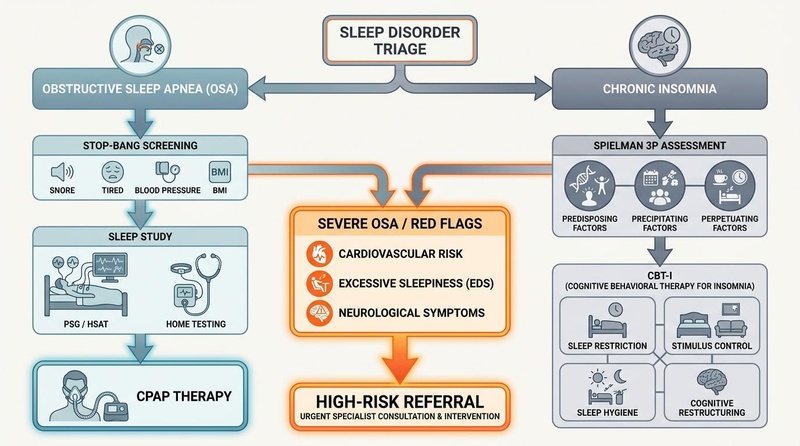
¶ Chronic Insomnia
Insomnia is defined by persistent difficulty with sleep initiation, duration, consolidation, or quality, despite adequate opportunity for sleep, resulting in daytime impairment[52]. It is clinically distinguished from acute sleep loss by duration, requiring symptoms to persist for at least 3 nights per week for a minimum of 3 months[52:1].
The development and maintenance of chronic insomnia is best understood through Spielman's 3P Model[53]:
- Predisposing Factors: Baseline biological or psychological traits that lower the threshold for sleep disturbance (e.g., genetic hyperarousal, anxiety traits, older age)[53:1][54].
- Precipitating Factors: Acute triggers that initiate sleep disruption (e.g., acute stress, medical illness, major life events)[53:2].
- Perpetuating Factors: Maladaptive behaviors and cognitive patterns adopted to cope with sleep loss, which subsequently maintain the insomnia after the precipitating factor has resolved (e.g., spending excessive time in bed, irregular wake times, pre-bed cognitive rumination)[54:1].
¶ Cognitive Behavioral Therapy for Insomnia (CBT-I)
CBT-I is the globally recommended first-line gold-standard treatment for chronic insomnia, demonstrating superior long-term efficacy and safety compared to pharmacotherapy[52:2][55]. CBT-I is a structured, multi-component psychological intervention targeting perpetuating factors:
- Stimulus Control Therapy: Re-establishes the bed and bedroom as strong cues for sleep rather than frustration and wakefulness. Rules include going to bed only when sleepy, leaving the bed if awake for >20 minutes, and maintaining a strict wake time[52:3].
- Sleep Restriction Therapy (SRT): Artificially limits the time spent in bed to closely match the patient's actual sleep duration, consolidating sleep. For example, if a patient spends 8 hours in bed but only sleeps 5 hours, SRT restricts their sleep window to 5.5 hours. This accumulates homeostatic sleep pressure (Process S), improving sleep efficiency before the window is gradually widened[56].
- Cognitive Restructuring: Addresses catastrophic beliefs and unrealistic expectations regarding sleep loss ("I will never function tomorrow if I don't sleep now"), reducing pre-bed hyperarousal[56:1].
- Relaxation Training: Teaches progressive muscle relaxation, autogenic training, or diaphragmatic breathing to damp down autonomic hyperarousal.
- Sleep Hygiene Education: Provides general guidelines on light, caffeine, alcohol, and bedroom environment to support sleep architecture.
¶ Obstructive Sleep Apnea (OSA)
Obstructive Sleep Apnea is a highly prevalent sleep-related breathing disorder characterized by repetitive partial (hypopnea) or complete (apnea) collapse of the upper airway during sleep, despite ongoing respiratory effort[57]. This mechanical collapse triggers cyclic cascades of:
- Intermittent Hypoxia: Repetitive drops in blood oxygen saturation, triggering oxidative stress and systemic vascular inflammation[58].
- Intrusion of Sleep Architecture: Brief micro-arousals required to restore airway muscle tone, severely fragmenting sleep and abolishing N3 and REM sleep.
- Autonomic Surges: Explosive sympathetic nervous system activation, driving acute spikes in blood pressure and heart rate during sleep[11:1].
¶ Clinical Screening & Triage: The STOP-BANG Score
A clinical score of 3 or higher on the validated STOP-BANG questionnaire indicates a moderate-to-high risk of OSA, requiring formal diagnostic triage:
| Letter | Metric | Criteria |
|---|---|---|
| S | Snoring | Do you snore loudly (louder than talking or heard through closed doors)? |
| T | Tiredness | Do you often feel tired, fatigued, or sleepy during the daytime? |
| O | Observed | Has anyone observed you stop breathing or choking/gasping during sleep? |
| P | Pressure | Do you have or are you being treated for high blood pressure? |
| B | Body Mass Index | Is your BMI greater than 35 ? |
| A | Age | Are you older than 50 years of age? |
| N | Neck Circumference | Is your neck circumference greater than 40 cm (15.7 inches)? |
| G | Gender | Are you male? |
Definitive diagnosis requires polysomnography (PSG) or a Home Sleep Apnea Test (HSAT). Treatment is critical: untreated severe OSA carries a 2.25-fold increased risk of cardiovascular death[11:2]. Continuous Positive Airway Pressure (CPAP) is the gold-standard therapeutic intervention, maintaining mechanical airway patency and completely resolving intermittent hypoxia and sleep fragmentation[12:1].
¶ Restless Legs Syndrome (RLS)
RLS is a sensorimotor disorder characterized by an irresistible urge to move the legs, typically accompanied by uncomfortable sensations. Symptoms follow a circadian pattern, worsening during evening or night hours of rest and temporarily resolving with movement. RLS is strongly linked to central dopamine dysfunction and brain iron deficiency[59]. Clinicians should evaluate serum ferritin levels in all RLS patients; maintaining ferritin levels above 75 ng/mL via oral or intravenous iron supplementation is a first-line clinical strategy to alleviate symptoms.
¶ REM Sleep Behavior Disorder (RBD) & Synucleinopathies
While many parasomnias (such as sleepwalking or night terrors) occur during NREM sleep and are typically benign[37:1], RBD is a REM-sleep parasomnia characterized by the loss of normal skeletal muscle atonia, leading patients to physically enact vivid, often violent dreams[15:1].
RBD is a profound clinical red flag. It serves as a highly specific prodromal marker for alpha-synucleinopathies (neurodegenerative disorders marked by toxic alpha-synuclein aggregates), including Parkinson’s disease, Dementia with Lewy Bodies (DLB), and Multiple System Atrophy (MSA)[15:2]. Longitudinal cohort studies demonstrate that up to 80% of patients diagnosed with idiopathic RBD will convert to a clinically defined synucleinopathy within 10 to 15 years of RBD symptom onset[15:3]. Immediate referral to a sleep specialist and neurologist is mandatory for patient protection and long-term neuroprotective planning.
¶ Practical Applications: Measurement & Wearables
The modern democratization of sleep tracking has enabled continuous, longitudinal monitoring of sleep patterns outside clinical labs. Understanding the capabilities and limitations of consumer wearables is critical for effective tracking.
| Metric Type | Technology | Clinical Efficacy | Use Case / Limitations |
|---|---|---|---|
| Sleep Regularity & Consistency | Actigraphy / Accelerometry | High | Superb for tracking day-to-day bedtime consistency, awakenings, and circadian stability[44:1]. |
| Total Sleep Duration | Actigraphy / PPG Fusion | High | Highly sensitive (≥95%) at detecting sleep-wake states and calculating total nocturnal duration[43:1]. |
| Autonomic Nervous System (ANS) State | Photoplethysmography (PPG) | High | Tracks resting heart rate and Heart Rate Variability (HRV) as indicators of somatic recovery[60]. |
| Sleep Stage Discrimination (N3 vs. REM) | Multisensor PPG & Accelerometry | Low to Moderate | Significantly less accurate at distinguishing N1, N2, N3, and REM stages (50–86% sensitivity)[45:1]. |
| Airway Patency / SpO2 | Pulse Oximetry | Moderate | Monitors overnight oxygen desaturation; useful for highlighting OSA risk but not diagnostic[61]. |
¶ Wearable Limitations & Skin Tone Discrepancies
Consumer sleep trackers are not diagnostic devices. Users must avoid "orthosomnia"—an unhealthy obsession with achieving perfect sleep tracker metrics that paradoxically exacerbates sleep-onset anxiety[61:1]. Additionally, photoplethysmography (PPG) sensors use green light to measure blood flow changes. Studies have validated that optical sensors exhibit lower accuracy and higher rates of signal dropout in individuals with darker skin tones, as melanin absorbs green light more efficiently[46:1]. For clinical diagnostic purposes, gold-standard polysomnography (PSG) remains the absolute standard.
¶ Evidence-Based Sleep Supplements
While behavioral and environmental optimization represent the primary therapy for sleep disorders, targeted supplemental compounds can support sleep architecture and physiological relaxation.
¶ 1. Magnesium Bisglycinate
- Dose: 200–400 mg pre-bed.
- Mechanism: Magnesium acts as an antagonist of the NMDA (glutamate) receptor and an agonist of GABAergic pathways, reducing central nervous system hyperexcitability and promoting somatic muscle relaxation[21:1].
- Evidence Quality: Moderate (RCTs demonstrate improved sleep efficiency and subjective quality)[21:2].
¶ 2. Glycine
- Dose: 3 grams pre-bed.
- Mechanism: Acts as an inhibitory neurotransmitter in the brainstem and spinal cord. It stimulates NMDA receptors in the suprachiasmatic nucleus, promoting cutaneous vasodilation in the hands and feet to accelerate core body temperature cooling[23:2].
- Evidence Quality: High (RCTs show reduced sleep latency, enhanced slow-wave sleep, and reduced next-day fatigue)[22:1].
¶ 3. L-Theanine
- Dose: 100–200 mg pre-bed.
- Mechanism: An amino acid analog that crosses the blood-brain barrier to inhibit cortical glutamate receptors and increase brain levels of GABA, dopamine, and glycine. It promotes alpha-wave brain activity, signaling alert relaxation[24:1].
- Evidence Quality: Moderate (RCTs show decreased sleep latency and reduced next-day subjective stress)[24:2].
¶ 4. Melatonin
- Dose: 0.3–3 mg (low-dose chronobiotic).
- Mechanism: Direct agonist of central melatonin and receptors in the SCN, serving as a phase-shifting agent (chronobiotic) to align the circadian clock[62][51:1].
- Evidence Quality: High for circadian phase adjustment and jet lag; Moderate for primary insomnia[62:1][63].
¶ 5. Apigenin
- Dose: 50–100 mg pre-bed.
- Mechanism: A bioflavonoid that acts as a positive allosteric modulator of central receptors (similar to, but weaker than, benzodiazepines), exerting mild sedative and anxiolytic properties.
- Evidence Quality: Low to Moderate (supported primarily by pilot trials and mechanistic pathways).
¶ 6. Inositol
- Dose: 1–2 grams pre-bed.
- Mechanism: Acts as a second messenger precursor in intracellular signaling pathways, specifically modulating serotonin and GABA receptor sensitivity to reduce nocturnal anxiety and facilitate relaxation.
- Evidence Quality: Low (mechanistic support with limited direct human clinical trials for sleep).
¶ Evidence Summary Table (Human Outcomes)
The following table summarizes key clinical and epidemiological outcomes associated with sleep duration, regularity, and targeted interventions in human populations:
| Outcome / Goal | Effect | Consistency | Evidence Quality | Trials | Notes (Population, Duration, Dose) |
|---|---|---|---|---|---|
| All-Cause Mortality (Short sleep <7h/night) | High | Moderate | 79 Cohort Studies | [14% increased mortality risk vs. 7–8h reference range][6:3] | |
| All-Cause Mortality (High sleep regularity) | High | Moderate | 60,000+ Participants | [20% to 48% lower mortality risk in highly consistent sleepers][5:2] | |
| Cardio-Metabolic Mortality (Sleep regularity) | High | Moderate | 60,000+ Participants | [22% to 57% lower cardiometabolic mortality in top regularity quintiles][5:3] | |
| Type 2 Diabetes Risk (Short sleep <6h/night) | High | Moderate | Meta-analysis | [28% increased risk of incident T2D vs. 6–8h baseline][9:1] | |
| Cardiovascular Mortality (Untreated Severe OSA) | High | Moderate | Large Prospective | [2.25-fold increased risk of death in severe sleep apnea][11:3] | |
| Subjective Sleep Quality (Glycine supplement) | High | Moderate | RCTs | [3 grams pre-bed reduces sleep latency and next-day fatigue][22:2] | |
| Sleep Onset Latency (Melatonin supplement) | High | Moderate | Meta-analysis | [Modestly decreases sleep latency by 4 to 8 minutes][62:2] | |
| Sleep Quality & Deep Sleep (Magnesium Bisglycinate) | Moderate | Moderate | Clinical Trials | [Supplemental magnesium improves subjective quality and deep sleep stages][64][21:3] | |
| Subjective Insomnia Severity (CBT-I therapy) | High | High | Clinical Guidelines | [Gold-standard non-pharmacological resolution of chronic insomnia][52:4][65] | |
| Cortical EEG Slow-Wave Density (Caffeine intake) | High | High | Clinical Trials | [150-300 mg caffeine disrupts slow-wave activity and alters nocturnal EEG power][20:2] | |
| Overnight Autonomic Recovery (Evening Alcohol) | High | Moderate | Large Cohorts | [Severe reduction in overnight HRV and dramatic elevation of resting heart rate][18:4] | |
| SWSD Daytime Sleep Latency (Melatonin 1-3 mg) | High | Moderate | Systematic Reviews | [Timed administration improves daytime sleep consolidation for night workers][51:2] |
Mechanisms of Action
Glymphatic System and Waste Clearance
During sleep, the brain activates the glymphatic system, a waste clearance pathway where cerebrospinal fluid (CSF) flows into paravascular spaces around cerebral arteries, mixes with interstitial fluid, and removes metabolic waste products[8:5][37:2]. The extracellular space increases by approximately 60% during sleep to promote clearance of interstitial wastes including amyloid beta (Aβ) and tau proteins associated with neurodegenerative diseases[66]. Slow wave sleep (deep sleep) plays a particularly important role in moving waste products out of the brain[41:1]. Even one night of sleep deprivation in young adults significantly increases amyloid beta accumulation in the hippocampus and thalamus[42:1].
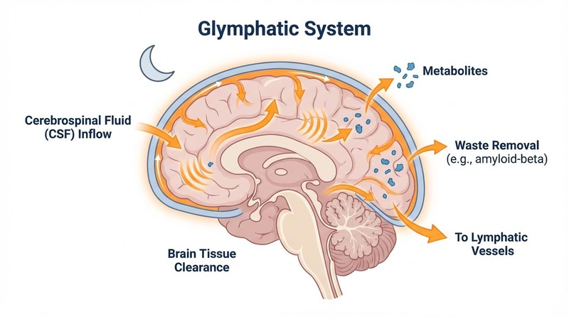
Figure 2: Sagittal diagram representing the glymphatic system. During slow-wave sleep, the interstitial space increases, enabling rhythmic oscillations of cerebrospinal fluid (CSF) to wash neurotoxic proteins from the brain parenchyma.
Autophagy and Cellular Repair
Sleep promotes autophagy, a cellular self-cleaning process where cells degrade and recycle damaged proteins and organelles[35:1][67]. During waking hours, proteotoxic compounds accumulate and are degraded by the proteasome during sleep[36:1]. The protein PARP1 senses mounting DNA damage and signals when sleep is needed; during sleep, DNA repair occurs efficiently through increased activity of DNA damage response proteins Rad52 and Ku80[34:2][32:2]. Sleep increases chromosome dynamics in neurons, which are necessary to reduce DNA double-strand breaks (DSBs) that accumulate during wakefulness, protecting against genomic instability[33:1].
Sleep fragmentation can dysregulate autophagy by blocking autophagy maturation processes, resulting in decreased autophagy flux and accumulation of cellular waste[68]. This relationship is particularly important for neuronal health, as neurons are not renewed and rely on efficient DNA repair mechanisms that function optimally during sleep[69].
Inflammation and Immune Function
Sleep exhibits bidirectional regulation with the immune system: insufficient sleep induces inflammatory responses, while immune activation affects sleep[70]. Sleep deprivation activates cellular markers of inflammation, increasing production of pro-inflammatory cytokines including interleukin-1β (IL-1β), tumor necrosis factor α (TNFα), interleukin-6 (IL-6), and C-reactive protein (CRP)[71][72]. Chronic sleep restriction results in low-grade inflammation that contributes to metabolic and neurodegenerative disorders[73].
In older adults, one marker of an aging immune system is increased basal levels of pro-inflammatory cytokines; IL-6 and TNF-α concentrations show over 2-fold increases in healthy older adults compared to young controls[74]. Adequate sleep quantity and quality support immune function, reduce infectious disease risk, and improve vaccination responses[75].
Metabolic and Hormonal Regulation
Sleep deprivation disrupts multiple metabolic hormones critical for health and longevity. Acute total sleep deprivation results in lower fasting serum concentrations of leptin (the satiety hormone) and higher plasma levels of ghrelin (the hunger hormone), likely increasing appetite and contributing to weight gain and metabolic dysfunction[38:1][39:1]. Growth hormone (GH) secretion occurs primarily during deep sleep stages, with sleep deprivation substantially dampening or abolishing the sleep-associated GH pulse[40:2].
Sleep restriction to less than 6 hours per night is associated with disturbances in glucose metabolism and insulin sensitivity, contributing to type 2 diabetes development[76]. The risk of developing type 2 diabetes in people with sustained sleep deprivation is comparable to risks from other well-known cardiometabolic risk factors[77].
Cardiovascular Function
Sleep deprivation disrupts autonomic nervous system balance, increasing sympathetic activity and leading to elevated heart rate, blood pressure, and reduced heart rate variability[10:1]. Experimental sleep deprivation leads to increased blood pressure and, in hospitalized patients, causes lack of nocturnal blood pressure dipping with persistently elevated sympathetic nervous system activity[78]. Sleep deprivation also impairs endothelial function (the health and responsiveness of blood vessel lining cells), resulting in reduced vasodilation capacity and increased vascular resistance[79].
Circadian Rhythm Regulation
The relationship between aging and circadian behavior is bidirectional: dysfunction of circadian clocks promotes age-related diseases, while aging leads to changes and disruption in circadian behavior and physiology[28:1]. Circadian rhythms play vital roles in health, and prolonged disruptions are associated with increased risks of type 2 diabetes, cancer, and cardiovascular disease[50:2]. Sleep/wake patterns change markedly with age, becoming increasingly fragmented in many cases[80]. Maintaining healthy circadian rhythms through appropriate light exposure and consistent sleep schedules may be crucial for healthy aging[81].
¶ References
Besedovsky L, Lange T, Haack M. The Sleep-Immune Crosstalk in Health and Disease. Physiological Reviews. 2019;99(3):1325-1380. https://journals.physiology.org/doi/full/10.1152/physrev.00010.2018 ↩︎
Xie L, Kang H, Xu Q, et al. Sleep drives metabolite clearance from the adult brain. Science. 2013;342(6156):373-377. https://www.science.org/doi/10.1126/science.1241224 ↩︎ ↩︎ ↩︎
Mander BA, Winer JR, Walker MP. Sleep and Human Aging. Neuron. 2017;94(1):19-36. https://www.cell.com/neuron/fulltext/S0896-6273(17)30088-2 ↩︎ ↩︎
Unraveling the interplay between sleep, redox metabolism, and aging: implications for brain health and longevity. Frontiers in Aging. 2025. https://www.frontiersin.org/journals/aging/articles/10.3389/fragi.2025.1605070/full ↩︎
Windred DP, Burns AC, Lane JM, et al. Sleep regularity is a stronger predictor of mortality risk than sleep duration: A prospective cohort study. SLEEP. 2024;47(1):zsad253. https://academic.oup.com/sleep/article/47/1/zsad253/7280269 ↩︎ ↩︎ ↩︎ ↩︎
Imbalanced sleep increases mortality risk by 14-34%: a meta-analysis. GeroScience. 2025. https://link.springer.com/article/10.1007/s11357-025-01592-y ↩︎ ↩︎ ↩︎ ↩︎
Grandner MA, Drummond SP. Who are the long sleepers? Towards an understanding of the mortality relationship. Sleep Medicine Reviews. 2007;11(5):341-360. https://www.sciencedirect.com/science/article/abs/pii/S1087079207000524 ↩︎
Jessen NA, Munk AS, Lundgaard I, Nedergaard M. The Glymphatic System: A Beginner's Guide. Neurochemical Research. 2015;40(12):2583-2599. https://link.springer.com/article/10.1007/s11064-015-1581-6 ↩︎ ↩︎ ↩︎ ↩︎ ↩︎ ↩︎
Shan Z, Ma H, Xie M, et al. Sleep duration and risk of type 2 diabetes: a meta-analysis of prospective studies. Diabetes Care. 2015;38(3):529-537. https://diabetesjournals.org/care/article/38/3/529/37749/Sleep-Duration-and-Risk-of-Type-2-Diabetes-A-Meta ↩︎ ↩︎
Tobaldini E, Costantino G, Solbiati M, et al. Sleep, sleep deprivation, autonomic nervous system and cardiovascular diseases. Neuroscience & Biobehavioral Reviews. 2017;74(Pt B):321-329. https://www.sciencedirect.com/science/article/abs/pii/S0149763416303682 ↩︎ ↩︎
Martinez-Garcia MA, Campos-Rodriguez F, Catalan-Serra P, et al. Cardiovascular Mortality in Obstructive Sleep Apnea in the Elderly. American Journal of Respiratory and Critical Care Medicine. 2012;186(9):909-916. https://www.atsjournals.org/doi/10.1164/rccm.201203-0448OC ↩︎ ↩︎ ↩︎ ↩︎
Marin JM, Carrizo SJ, Vicente E, Agusti AG. Long-term cardiovascular outcomes in men with obstructive sleep apnoea-hypopnoea with or without treatment with continuous positive airway pressure. The Lancet. 2005;365(9464):1046-1053. https://www.thelancet.com/journals/lancet/article/PIIS0140-6736(05)71141-7/fulltext ↩︎ ↩︎
Glass J, Lanctôt KL, Herrmann N, Sproule BA, Busto UE. Sedative hypnotics in older people with insomnia: meta-analysis of risks and benefits. BMJ. 2005;331(7526):1169. https://www.bmj.com/content/331/7526/1169 ↩︎
Glass J, Lanctôt KL, Herrmann N, Sproule BA, Busto UE. Sedative hypnotics in older people with insomnia: meta-analysis of risks and benefits. BMJ. 2005;331(7526):1169. https://www.bmj.com/content/331/7526/1169 ↩︎
Bellesi M, Castelnovo A. Why do we sleepwalk? A noradrenergic hypothesis of NREM sleep parasomnias. Sleep Medicine Reviews. 2026;76:101832. https://pubmed.ncbi.nlm.nih.gov/41932139/ ↩︎ ↩︎ ↩︎ ↩︎
Blue light has a dark side. Harvard Health Publishing. 2022. https://www.health.harvard.edu/staying-healthy/blue-light-has-a-dark-side ↩︎
Irish LA, Kline CE, Gunn HE, Buysse DJ, Hall MH. The role of sleep hygiene in promoting public health. Sleep Medicine Reviews. 2015;22:23-36. https://www.sciencedirect.com/science/article/abs/pii/S1087079214000811 ↩︎ ↩︎ ↩︎ ↩︎
Grosicki GJ, Robinson AT, Joyner MJ. Real-world effects of alcohol on heart rate, sleep, and physical activity by age and sex. PLOS Digital Health. 2026;5(3):e0000452. https://pubmed.ncbi.nlm.nih.gov/41801993/ ↩︎ ↩︎ ↩︎ ↩︎ ↩︎
Chang AM, Aeschbach D, Duffy JF, Czeisler CA. Evening use of light-emitting eReaders negatively affects sleep, circadian timing, and next-morning alertness. PNAS. 2015;112(4):1232-1237. https://www.pnas.org/doi/full/10.1073/pnas.1418490112 ↩︎ ↩︎ ↩︎
Chmiel J, Kurpas D. The Caffeinated Brain Part 2: The Effect of Caffeine on Sleep-Related Electroencephalography (EEG)-A Systematic and Mechanistic Review. Nutrients. 2026;18(8):1024. https://pubmed.ncbi.nlm.nih.gov/42075032/ ↩︎ ↩︎ ↩︎
Magnesium Bisglycinate Supplementation in Healthy Adults Reporting Poor Sleep: A Randomized, Placebo-Controlled Trial. Nutrition and Healthy Aging. 2024. https://pmc.ncbi.nlm.nih.gov/articles/PMC12412596/ ↩︎ ↩︎ ↩︎ ↩︎
Bannai M, et al. The effects of glycine on subjective daytime performance in partially sleep-restricted healthy volunteers. Frontiers in Neurology. 2012;3:61. https://pmc.ncbi.nlm.nih.gov/articles/PMC3328957/ ↩︎ ↩︎ ↩︎
Kawai N, et al. The sleep-promoting and hypothermic effects of glycine are mediated by NMDA receptors in the suprachiasmatic nucleus. Neuropsychopharmacology. 2015;40(6):1405-1416. https://pmc.ncbi.nlm.nih.gov/articles/PMC4397399/ ↩︎ ↩︎ ↩︎
Hidese S, et al. Effects of L-Theanine Administration on Stress-Related Symptoms and Cognitive Functions in Healthy Adults. Nutrients. 2019;11(10):2362. https://pubmed.ncbi.nlm.nih.gov/31623400/ ↩︎ ↩︎ ↩︎
Patel AK, Reddy V, Shumway KR, Araujo JF. Physiology, Sleep Stages. StatPearls. 2023. https://www.ncbi.nlm.nih.gov/books/NBK526132/ ↩︎ ↩︎ ↩︎ ↩︎
Carskadon MA, Dement WC. Normal Human Sleep: An Overview. Principles and Practice of Sleep Medicine. 2011:16-26. https://www.sciencedirect.com/science/article/abs/pii/B9781416066453000024 ↩︎ ↩︎
Hood S, Amir S. The aging clock: circadian rhythms and later life. Journal of Clinical Investigation. 2017;127(2):437-446. https://pmc.ncbi.nlm.nih.gov/articles/PMC5272178/ ↩︎
Kondratova AA, Kondratov RV. The circadian clock and pathology of the ageing brain. Nature Reviews Neuroscience. 2012;13(5):325-335. https://www.nature.com/articles/nrn3208 ↩︎ ↩︎
Shi SQ, Ansari TS, McGuinness OP, Wasserman DH, Johnson CH. Circadian disruption leads to insulin resistance and obesity. Current Biology. 2013;23(5):372-381. https://www.cell.com/current-biology/fulltext/S0960-9822(13)00126-8 ↩︎ ↩︎
Xie Y, Tang Q, Chen G, et al. New insights into the circadian rhythm and its related diseases. Frontiers in Physiology. 2019;10:682. https://www.frontiersin.org/articles/10.3389/fphys.2019.00682/full ↩︎ ↩︎
Tosini G, Ferguson I, Tsubota K. Effects of blue light on the circadian system and eye physiology. Molecular Vision. 2016;22:61-72. https://www.ncbi.nlm.nih.gov/pmc/articles/PMC4734149/ ↩︎ ↩︎ ↩︎ ↩︎
Thakkar N, Ramakrishnan A, Zada D, et al. Parp1 promotes sleep, which enhances DNA repair in neurons. Molecular Cell. 2021;81(24):4979-4993. https://pmc.ncbi.nlm.nih.gov/articles/PMC8688325/ ↩︎ ↩︎ ↩︎
Sleep: DNA Repair Function for Better Neuronal Aging? Current Biology. 2019;29(12):R557-R559. https://www.cell.com/current-biology/fulltext/S0960-9822(19)30551-2 ↩︎ ↩︎
Zada D, Bronshtein I, Lerer-Goldshtein T, et al. Sleep increases chromosome dynamics to enable reduction of accumulating DNA damage in single neurons. Nature Communications. 2019;10:895. https://www.nature.com/articles/s41467-019-08806-w ↩︎ ↩︎ ↩︎
Rest, Repair, Repeat: The Complex Relationship of Autophagy and Sleep. Journal of Molecular Biology. 2025. https://www.sciencedirect.com/science/article/pii/S0022283625002931 ↩︎ ↩︎
Impact of Sleep on Autophagy and Neurodegenerative Disease. Scientific Archives. 2024. https://www.scientificarchives.com/article/impact-of-sleep-on-autophagy-and-neurodegenerative-disease-sleeping-your-mind-clear ↩︎ ↩︎
The Sleeping Brain: Harnessing the Power of the Glymphatic System through Lifestyle Choices. Brain Sciences. 2020. https://pmc.ncbi.nlm.nih.gov/articles/PMC7698404/ ↩︎ ↩︎ ↩︎
Spiegel K, Tasali E, Leproult R, Van Cauter E. Effects of poor and short sleep on glucose metabolism and obesity risk. Nature Reviews Endocrinology. 2009;5(5):253-261. https://www.nature.com/articles/nrendo.2009.23 ↩︎ ↩︎
Spiegel K, Tasali E, Penev P, Van Cauter E. Brief communication: Sleep curtailment in healthy young men is associated with decreased leptin levels, elevated ghrelin levels, and increased hunger and appetite. Annals of Internal Medicine. 2004;141(11):846-850. https://journals.plos.org/plosmedicine/article?id=10.1371/journal.pmed.0010062 ↩︎ ↩︎
Copinschi G, Leproult R, Spiegel K. The important role of sleep in metabolism. Frontiers of Hormone Research. 2014;42:59-72. https://karger.com/books/book/421/chapter/5530967/The-Important-Role-of-Sleep-in-Metabolism ↩︎ ↩︎ ↩︎
Fultz NE, Bonmassar G, Setsompop K, et al. Coupled electrophysiological, hemodynamic, and cerebrospinal fluid oscillations in human sleep. Science. 2019;366(6465):628-631. https://www.science.org/doi/10.1126/science.aax5440 ↩︎ ↩︎
Shokri-Kojori E, Wang GJ, Wiers CE, et al. β-Amyloid accumulation in the human brain after one night of sleep deprivation. PNAS. 2018;115(17):4483-4488. https://pmc.ncbi.nlm.nih.gov/articles/PMC5924922/ ↩︎ ↩︎
Miller DJ, Sargent C, Roach GD. Accuracy of 11 Wearable, Nearable, and Airable Consumer Sleep Trackers: Prospective Multicenter Validation Study. JMIR mHealth and uHealth. 2023;11:e50983. https://pmc.ncbi.nlm.nih.gov/articles/PMC10654909/ ↩︎ ↩︎
Chinoy ED, Cuellar JA, Huwa KE, et al. Performance of seven consumer sleep-tracking devices compared with polysomnography. Sleep. 2021;44(5):zsaa291. https://pmc.ncbi.nlm.nih.gov/articles/PMC8120339/ ↩︎ ↩︎
Miller DJ, Sargent C, Roach GD. Accuracy of 11 Wearable, Nearable, and Airable Consumer Sleep Trackers: Prospective Multicenter Validation Study. JMIR mHealth and uHealth. 2023;11:e50983. https://pmc.ncbi.nlm.nih.gov/articles/PMC10654909/ ↩︎ ↩︎
Bent B, Goldstein BA, Kibbe WA, Dunn JP. Investigating sources of inaccuracy in wearable optical heart rate sensors. npj Digital Medicine. 2020;3:18. https://www.nature.com/articles/s41746-020-0226-6 ↩︎ ↩︎
Pierzchała K, Bielska W, Boczar Z. Thermoregulation in Sleep Disorders-Comprehensive Review. Journal of Clinical Medicine. 2026;15(7):1102. https://pubmed.ncbi.nlm.nih.gov/42074731/ ↩︎ ↩︎ ↩︎ ↩︎
Andersen ML, Hachul H, Ishikura IA. Sleep in women: a narrative review of hormonal influences, sex differences and health implications. Frontiers in Sleep. 2023;2:1025542. https://pubmed.ncbi.nlm.nih.gov/41426438/ ↩︎ ↩︎ ↩︎ ↩︎ ↩︎ ↩︎
Ben Jemia E, Trimech S, Saidani A. Diagnostic challenges of obstructive sleep apnea syndrome: A comparative study between elderly and younger populations. La Tunisie Medicale. 2026;104(3):142-149. https://pubmed.ncbi.nlm.nih.gov/42246712/ ↩︎
Lunn RM, Blask DE, Coogan AN, et al. Health consequences of electric lighting practices in the modern world: A report on the National Toxicology Program's workshop on shift work at night, artificial light at night, and circadian disruption. Science of The Total Environment. 2017;607-608:1073-1084. https://www.sciencedirect.com/science/article/pii/S0048969717314729 ↩︎ ↩︎ ↩︎
Carriedo-Diez B, Tosoratto-Venturi JL, Cantón-Manzano C. The Effects of the Exogenous Melatonin on Shift Work Sleep Disorder in Health Personnel: A Systematic Review. International Journal of Environmental Research and Public Health. 2022;19(16):10112. https://pubmed.ncbi.nlm.nih.gov/36011832/ ↩︎ ↩︎ ↩︎
Edinger JD, Arnedt JT, Bertisch SM, et al. Behavioral and psychological treatments for chronic insomnia disorder in adults: an American Academy of Sleep Medicine clinical practice guideline. Journal of Clinical Sleep Medicine. 2021;17(2):255-262. https://jcsm.aasm.org/doi/10.5664/jcsm.8986 ↩︎ ↩︎ ↩︎ ↩︎ ↩︎
Moon S, You SY. Prevalence and risk factors of insomnia among older adults living alone: A cross-sectional study based on Spielman's 3P behavioral model. Geriatric Nursing. 2025;55(6):120-128. https://pubmed.ncbi.nlm.nih.gov/40946404/ ↩︎ ↩︎ ↩︎
Han EK, Son HK. The Factors Affecting Sleep Quality in Community-Dwelling Older Adults, Based on Spielman's 3P Model of Insomnia. Iranian Journal of Public Health. 2024;53(12):2640-2649. https://pubmed.ncbi.nlm.nih.gov/39759194/ ↩︎ ↩︎
Qaseem A, Kansagara D, Forciea MA, et al. Management of Chronic Insomnia Disorder in Adults: A Clinical Practice Guideline From the American College of Physicians. Annals of Internal Medicine. 2016;165(2):125-133. https://pubmed.ncbi.nlm.nih.gov/27136449/ ↩︎
Brasure M, Fuchs E, MacDonald R, et al. Psychological and Behavioral Interventions for Managing Insomnia Disorder: An Evidence Report for a Clinical Practice Guideline by the American College of Physicians. Annals of Internal Medicine. 2016;165(2):113-124. https://pubmed.ncbi.nlm.nih.gov/27136619/ ↩︎ ↩︎
Gottlieb DJ, Punjabi NM. Diagnosis and Management of Obstructive Sleep Apnea: A Review. JAMA. 2020;323(14):1389-1400. https://jamanetwork.com/journals/jama/fullarticle/2764461 ↩︎
Martínez-García MA, Campos-Rodríguez F, Barbé F. Cancer and OSA: An Ongoing Journey. Archivos de Bronconeumología. 2022;58(4):337-338. https://www.archbronconeumol.org/en-cancer-osa-an-ongoing-journey-articulo-S1579212921001935 ↩︎
Watson NF, Badr MS, Belenky G, et al. Recommended Amount of Sleep for a Healthy Adult: A Joint Consensus Statement of the American Academy of Sleep Medicine and Sleep Research Society. Sleep. 2015;38(6):843-844. https://academic.oup.com/sleep/article/38/6/843/2416980 ↩︎
Khosla S, Deak MC, Gault D, et al. Consumer sleep technology: an American Academy of Sleep Medicine position statement. Journal of Clinical Sleep Medicine. 2018;14(5):877-880. https://jcsm.aasm.org/doi/10.5664/jcsm.7128 ↩︎
Sleep tracking: A systematic review of the research using commercially available technology. Sleep Health. 2020;6(6):e94-e106. https://pmc.ncbi.nlm.nih.gov/articles/PMC7597680/ ↩︎ ↩︎
Ferracioli-Oda E, Qawasmi A, Bloch MH. Meta-analysis: melatonin for the treatment of primary sleep disorders. PLOS ONE. 2013;8(5):e63773. https://journals.plos.org/plosone/article?id=10.1371/journal.pone.0063773 ↩︎ ↩︎ ↩︎
Melatonin and your sleep. UC Davis Health. 2025. https://health.ucdavis.edu/blog/cultivating-health/melatonin-and-your-sleep-is-it-safe-what-are-the-side-effects-and-how-does-it-work/2025/02 ↩︎
Abbasi B, et al. The effect of magnesium supplementation on primary insomnia in elderly: a double-blind placebo-controlled clinical trial. Journal of Research in Medical Sciences. 2012;17(12):1161-1169. https://pubmed.ncbi.nlm.nih.gov/23853635/ ↩︎
Morin CM, Bootzin RR, Buysse DJ, Edinger JD, Espie CA, Lichstein KL. Psychological and behavioral treatment of insomnia: update of the recent evidence. Sleep. 2006;29(11):1398-1414. https://academic.oup.com/sleep/article/29/11/1398/2708120 ↩︎
Sleep and the glymphatic system. American Nurse. 2024. https://www.myamericannurse.com/sleep-and-the-glymphatic-system/ ↩︎
Mitochondrial autophagy in the sleeping brain. Frontiers in Cell and Developmental Biology. 2022. https://www.frontiersin.org/journals/cell-and-developmental-biology/articles/10.3389/fcell.2022.956394/full ↩︎
Naidoo N, Ferber M, Galante RJ, et al. Short-Term Sleep Fragmentation Dysregulates Autophagy in a Brain Region-Specific Manner. Frontiers in Neuroscience. 2021. https://pmc.ncbi.nlm.nih.gov/articles/PMC8538758/ ↩︎
Zada D, Tovin A, Lerer-Goldshtein T, Appelbaum L. Contribution of sleep to the repair of neuronal DNA double-strand breaks. Scientific Reports. 2016;6:36804. https://www.nature.com/articles/srep36804 ↩︎
Besedovsky L, Lange T, Born J. Sleep and immune function. Pflügers Archiv - European Journal of Physiology. 2012;463(1):121-137. https://link.springer.com/article/10.1007/s00424-011-1044-0 ↩︎
Irwin MR, Olmstead R, Carroll JE. Sleep Disturbance, Sleep Duration, and Inflammation: A Systematic Review and Meta-Analysis of Cohort Studies and Experimental Sleep Deprivation. Biological Psychiatry. 2016;80(1):40-52. https://www.biologicalpsychiatryjournal.com/article/S0006-3223(15)00437-0/fulltext ↩︎
Mullington JM, Simpson NS, Meier-Ewert HK, Haack M. Sleep loss and inflammation. Best Practice & Research Clinical Endocrinology & Metabolism. 2010;24(5):775-784. https://www.sciencedirect.com/science/article/abs/pii/S1521690X10000768 ↩︎
Sleep and Immune System Crosstalk: Implications for Inflammatory Homeostasis and Disease Pathogenesis. International Journal of Molecular Sciences. 2024. https://pmc.ncbi.nlm.nih.gov/articles/PMC11559494/ ↩︎
Opp MR, George A, Ringgold KM, Hansen KM, Bullock KM, Banks WA. Sleep and immune function: glial contributions and consequences of aging. Current Opinion in Neurobiology. 2013;23(5):806-811. https://pmc.ncbi.nlm.nih.gov/articles/PMC3695049/ ↩︎
Besedovsky L, Lange T, Haack M. The Sleep-Immune Crosstalk in Health and Disease. Physiological Reviews. 2019;99(3):1325-1380. https://journals.physiology.org/doi/full/10.1152/physrev.00010.2018 ↩︎
Knutson KL, Van Cauter E, Zee P, Liu K, Lauderdale DS. Cross-sectional associations between measures of sleep and markers of glucose metabolism among subjects with and without diabetes. Diabetes Care. 2011;34(5):1171-1176. https://diabetesjournals.org/care/article/34/5/1171/38538/Cross-Sectional-Associations-Between-Measures-of ↩︎
Cardiovascular, Inflammatory and Metabolic Consequences of Sleep Deprivation. Progress in Cardiovascular Diseases. 2009;51(4):294-302. https://pmc.ncbi.nlm.nih.gov/articles/PMC3403737/ ↩︎
Kato M, Phillips BG, Sigurdsson G, Narkiewicz K, Pesek CA, Somers VK. Effects of Sleep Deprivation on Neural Circulatory Control. Hypertension. 2000;35(5):1173-1175. https://www.ahajournals.org/doi/10.1161/01.HYP.35.5.1173 ↩︎
Sauvet F, Leftheriotis G, Gomez-Merino D, et al. Effect of acute sleep deprivation on vascular function in healthy subjects. Journal of Applied Physiology. 2010;108(1):68-75. https://journals.physiology.org/doi/full/10.1152/japplphysiol.00851.2009 ↩︎
Mander BA, Winer JR, Walker MP. Sleep and Human Aging. Neuron. 2017;94(1):19-36. https://www.cell.com/neuron/fulltext/S0896-6273(17)30088-2 ↩︎
Huang YL, Liu RY, Wang QS, Van Someren EJ, Xu H, Zhou JN. Age-associated difference in circadian sleep-wake and rest-activity rhythms. Physiology & Behavior. 2002;76(4-5):597-603. https://www.sciencedirect.com/science/article/abs/pii/S0031938402007334 ↩︎